Role of Peanut Agglutinin in Cancer Detection
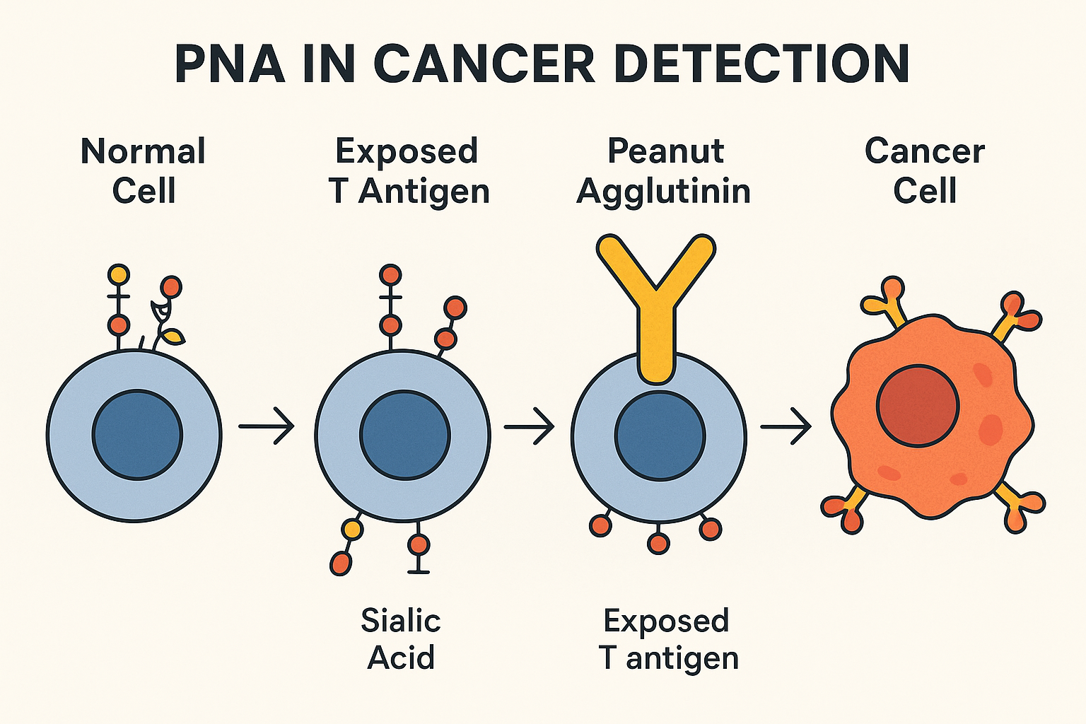
Peanut agglutinin (PNA) plays a role in cancer detection because it specifically binds to the Thomsen–Friedenreich antigen (T-antigen) — a carbohydrate structure that is hidden in normal cells but exposed in many cancer cells due to abnormal glycosylation.
How It Works in Cancer Detection
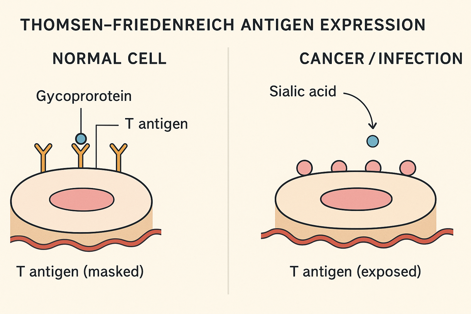
- Normal cells
- The T-antigen (Gal-β(1–3)-GalNAc) on cell-surface glycoproteins is usually capped with sialic acid.
- This “cap” hides the T-antigen from PNA binding.
- Cancer cells
- Many tumors (breast, colon, prostate, pancreas, lung, etc.) have incomplete glycosylation, meaning the sialic acid cap is missing.
- This exposes the T-antigen on the cell surface.
- PNA binds strongly to these exposed T-antigens.
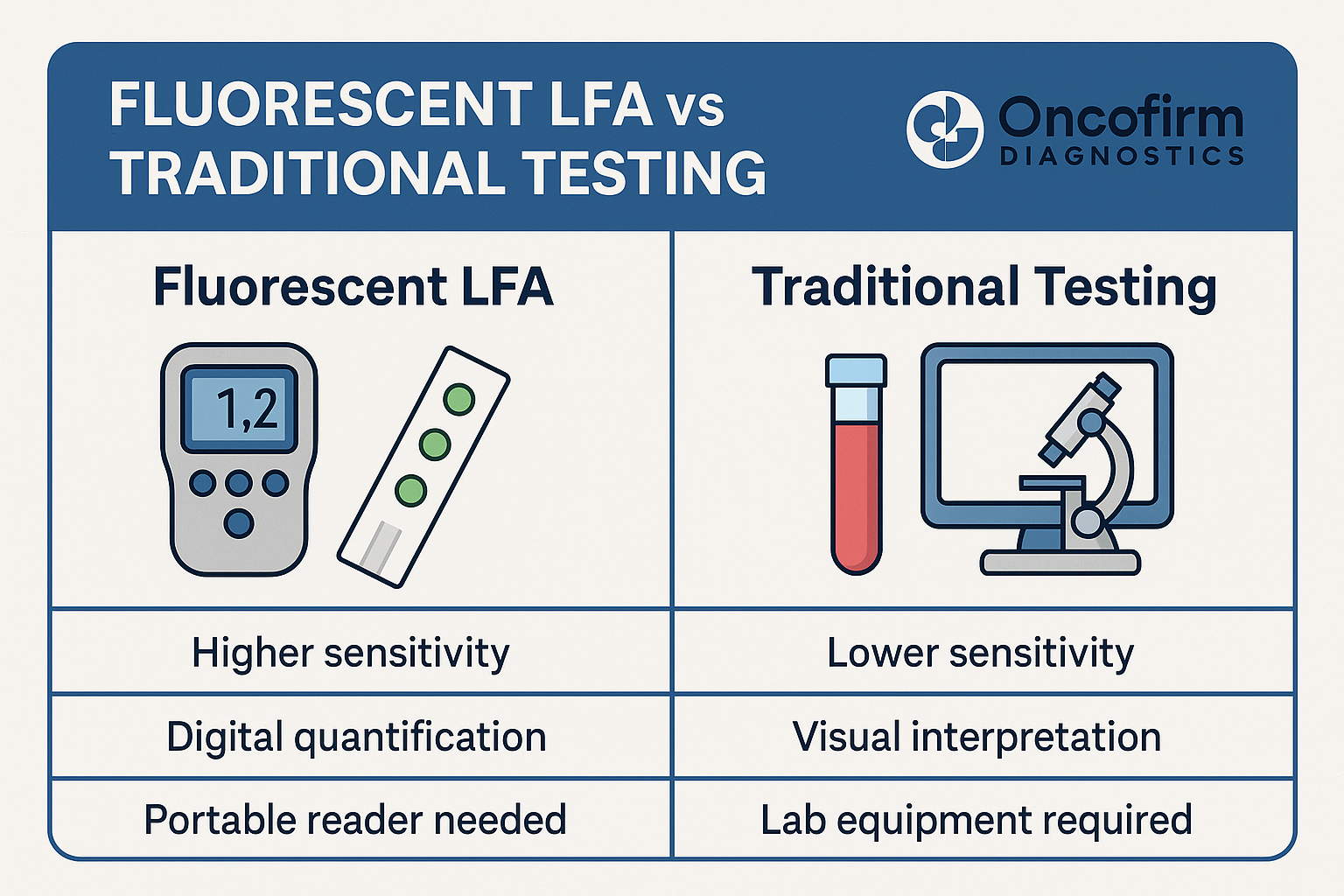
- Detection
- PNA can be tagged with fluorescent dyes, enzymes (for color reactions), or other markers.
- When applied to tissue sections (histochemistry) or cell samples, PNA will highlight cells with exposed T-antigen.
- Pathologists and researchers can then see which cells have this cancer-associated marker.
Why It’s Useful in Oncology
- Tumor marker: T-antigen exposure detected by PNA is correlated with tumor progression, invasiveness, and metastasis.
- Histopathology tool: Helps distinguish between malignant and benign tissues in research settings.
- Potential prognostic value: Higher PNA binding often relates to more aggressive cancers.