AI-Powered Multi-Biomarker Blood Tests for Cancer Screening and Triage | OncoFirm
Artificial Intelligence–Enabled Multi-Biomarker Blood Tests for Cancer Screening and Clinical Triage
Biological Rationale, Computational Architecture, Validation Requirements, and a Translational Framework for Point-of-Care Deployment
OncoFirm Diagnostics Scientific Affairs
Article type: Technical White Paper and Narrative Review
Publication date: July 2026
Correspondence: [email protected]
Abstract
Background
Blood-based cancer testing is advancing from the measurement of isolated tumor markers toward the integrated analysis of multiple molecular, cellular, immunologic, and physiological signals. Artificial intelligence and machine-learning methods may support this transition by identifying multivariable patterns that are not apparent when biomarkers are interpreted independently. However, the clinical value of an AI-enabled multi-biomarker test depends on its intended use, analytical reliability, calibration, population-specific performance, downstream diagnostic consequences, and demonstrated clinical utility.
Objective
This technical review describes the biological rationale, computational architecture, clinical applications, validation requirements, and governance principles relevant to AI-enabled multi-biomarker blood tests developed for cancer screening or clinical triage. It also presents a cautious translational framework applicable to the development of a digitally interpreted, multiplex point-of-care platform.
Methods
A narrative synthesis of established concepts in cancer biomarker science, diagnostic medicine, clinical prediction modeling, in vitro diagnostic development, and medical-device artificial intelligence was performed. This paper does not report new patient data, pooled performance estimates, or a systematic meta-analysis.
Findings
Multi-biomarker testing may integrate complementary information from circulating nucleic acids, proteins, antigens, autoantibodies, extracellular vesicles, metabolites, hematologic measurements, biochemical measurements, and patient characteristics. AI can support signal normalization, feature selection, multimodal integration, risk estimation, quality control, and uncertainty assessment. Nevertheless, models developed in retrospective case-control datasets may substantially overestimate performance when transferred to asymptomatic screening populations or heterogeneous clinical referral pathways. Clinical validation must therefore be conducted in the intended-use population using prespecified thresholds, independent datasets, stage- and cancer-specific analyses, appropriate reference standards, and longitudinal follow-up.
Conclusions
AI-enabled multi-biomarker blood testing is a scientifically plausible approach to cancer-risk assessment, but technical feasibility and high discrimination are not equivalent to clinical benefit. Population screening and symptomatic triage are separate medical uses requiring different thresholds, study designs, endpoints, and risk-management strategies. Future systems should be positioned as adjunctive clinical decision-support tools unless prospective evidence demonstrates that their use improves meaningful patient outcomes without introducing disproportionate diagnostic harm.
Keywords: artificial intelligence, machine learning, multi-biomarker blood test, cancer screening, clinical triage, early cancer detection, liquid biopsy, cancer biomarkers, fluorescent lateral flow assay, digital diagnostic reader, clinical decision support, in vitro diagnostics
1. Introduction
Cancer is biologically heterogeneous. Tumors arising in different organs—and molecular subtypes within the same organ—may release substantially different quantities and types of biological material into the circulation. Early-stage tumors may produce only weak or intermittent systemic signals, while nonmalignant inflammation, infection, tissue injury, age-related changes, benign neoplasia, and chronic disease may alter many of the same analytes.
This biological overlap limits the utility of relying on a single circulating biomarker as a universal indicator of malignancy. A multi-biomarker strategy attempts to address this limitation by evaluating several partially independent signals simultaneously. The underlying premise is that a pattern distributed across multiple biomarkers may contain more diagnostic information than any single component.
Artificial intelligence is relevant because the relationship between these biomarkers and cancer risk may be nonlinear, context-dependent, and influenced by interactions among patient characteristics, preanalytical variables, assay measurements, and disease biology. Machine-learning methods can estimate these relationships and convert multiple measurements into a calibrated probability or risk category.
A cancer screening test, however, does not establish a diagnosis. It identifies a signal or risk state that may justify further evaluation. Positive results generally require conventional diagnostic procedures, which may include imaging, endoscopy, repeat laboratory testing, specialist evaluation, or tissue biopsy. Similarly, a negative blood test cannot exclude every cancer or justify disregarding persistent clinical symptoms. Current standard-of-care cancer screening should continue according to applicable professional guidance, regardless of the result of an investigational multi-cancer blood test [R1, R14]. The National Cancer Institute similarly distinguishes cancer screening from diagnosis and emphasizes that multi-cancer detection tests predict the possible presence of cancer rather than establish it.
2. Terminology and Intended Use
2.1 Multi-biomarker testing
A multi-biomarker blood test measures or derives information from more than one biological variable. The biomarkers may originate from a single analytical modality, such as a panel of circulating proteins, or from multiple modalities, such as cell-free DNA methylation, protein concentrations, hematologic indices, and demographic variables.
“Multi-biomarker” does not necessarily mean “multi-cancer.” A multi-biomarker model may be developed to estimate the probability of one cancer, a defined group of cancers, or cancer of any type. These intended uses should not be treated as interchangeable.
2.2 Single-cancer and multi-cancer detection
A single-cancer detection test is intended to identify a signal associated with one specified malignancy. A multi-cancer detection test attempts to identify signals associated with more than one cancer type, frequently using an additional classifier to estimate the probable cancer signal of origin.
The ability to detect a general cancer-associated signal and the ability to identify its probable anatomical origin are separate performance claims. Each requires independent evaluation [R1].
2.3 Screening
Screening is performed in individuals who do not have recognized symptoms of the cancer being assessed. Disease prevalence is generally low, particularly for any individual cancer type. Consequently, even a relatively small false-positive rate can lead to substantial numbers of unnecessary investigations.
A screening test must be evaluated as part of a complete clinical pathway, including:
- Who is eligible for testing;
- How frequently testing occurs;
- What constitutes a positive, negative, or indeterminate result;
- Which diagnostic workup follows a positive result;
- How unresolved positive results are managed;
- Whether patients continue established cancer screening;
- Whether screening reduces advanced disease or cancer mortality;
- The physical, psychological, and economic harms of testing.
2.4 Clinical triage
Clinical triage is performed after a patient has entered a diagnostic pathway, usually because of symptoms, an abnormal examination, an imaging finding, an existing laboratory abnormality, or an elevated baseline risk. The objective is not population screening; it is to estimate the urgency or probability of disease within a clinically selected group.
An AI-enabled blood test used for triage may help prioritize patients for accelerated investigation, identify patients who require additional evaluation, or support carefully defined de-escalation pathways. Machine-learning models using routine blood measurements have been developed for risk stratification in symptomatic patients referred from primary care, illustrating the distinction between referral triage and general-population screening [R6]. Such models still require prospective evaluation within the actual pathway in which they will be used.
Table 1. Screening and triage are distinct intended uses
| Characteristic | Population screening | Clinical triage |
|---|---|---|
| Target population | Generally asymptomatic individuals | Symptomatic, referred, high-risk, or previously abnormal population |
| Pretest probability | Usually low | Usually higher and pathway dependent |
| Primary purpose | Detect a cancer-associated signal before clinical presentation | Prioritize, stratify, or guide further diagnostic evaluation |
| Principal performance concern | Very high specificity while maintaining clinically useful sensitivity | Sensitivity, calibration, safety of de-escalation, and pathway efficiency |
| Typical output | Screening signal detected or not detected; possibly predicted origin | Calibrated risk estimate or defined clinical risk category |
| Main potential harm | False positives, overdiagnosis, unnecessary invasive testing | Missed cancer, delayed diagnosis, or inappropriate pathway diversion |
| Required validation | Prospective screening population with longitudinal follow-up | Intended referral population under real-world workflow conditions |
| Relevant clinical-utility endpoint | Advanced-stage incidence, cancer mortality, harms, diagnostic burden | Time to diagnosis, missed-cancer rate, referral prioritization, resource utilization |
A model validated for one intended use must not be assumed to perform adequately in the other. Differences in prevalence, disease spectrum, stage distribution, comorbidity, and referral selection can materially alter calibration and predictive value.
3. Biological Components of Multi-Biomarker Blood Tests
3.1 Circulating tumor DNA and cell-free DNA
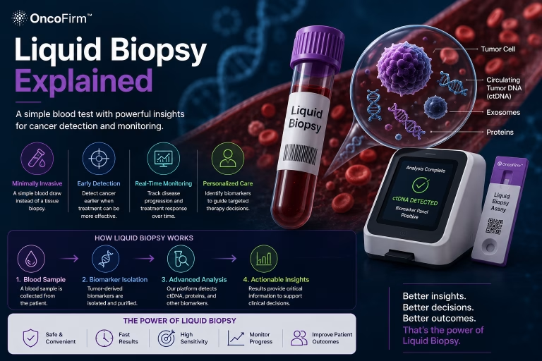
Cell-free DNA is released into the circulation from normal and abnormal tissues. The tumor-derived fraction, commonly described as circulating tumor DNA, may contain somatic mutations, copy-number alterations, methylation patterns, nucleosome footprints, or fragmentation characteristics associated with malignancy.
Mutation-based approaches may offer biological specificity but can be limited by low tumor fraction in early disease and by nonmalignant somatic variants, including variants arising from clonal hematopoiesis. Methylation and fragmentation approaches evaluate broader epigenetic or physical characteristics of cell-free DNA and may provide tissue-associated information.
Primary studies have demonstrated the technical feasibility of mutation-plus-protein assays, targeted methylation classifiers, and genome-wide fragmentation analysis for cancer signal detection [R2–R4]. These investigations establish scientific plausibility but do not, by themselves, demonstrate population-level clinical benefit.
3.2 Circulating RNA
Circulating RNA measurements may include messenger RNA, microRNA, long noncoding RNA, transfer-RNA fragments, and RNA associated with extracellular vesicles. RNA profiles can reflect both tumor biology and host response.
Potential limitations include molecular instability, sensitivity to collection and processing conditions, variation in normalization methods, and the possibility that inflammatory or tissue-injury responses may resemble cancer-associated patterns [R1].
3.3 Proteins and tumor-associated antigens
Circulating proteins and antigens are attractive for decentralized testing because they can often be measured using immunoassay technologies. Examples include established organ-associated tumor markers, inflammatory mediators, growth factors, glycoproteins, enzymes, and tumor-associated carbohydrate antigens.
Many protein biomarkers lack sufficient specificity when interpreted alone. Concentrations may be influenced by benign prostatic enlargement, hepatic dysfunction, renal dysfunction, infection, smoking, pregnancy, autoimmune disease, tissue injury, or other nonmalignant conditions. A multi-protein model may improve discrimination by considering relationships among several markers, but it can also learn confounding patterns if the development dataset is not representative.
3.4 Autoantibodies and immune-response biomarkers
The immune system may generate antibodies against altered, overexpressed, mislocalized, or aberrantly glycosylated tumor-associated molecules. Because an immune response may amplify a biologically small tumor signal, autoantibody panels are of interest for early detection.
However, autoantibody responses vary among patients and can overlap with autoimmune, infectious, and inflammatory conditions. Validation cohorts should therefore include clinically relevant nonmalignant controls rather than only healthy volunteers [R1].
3.5 Extracellular vesicles, circulating cells, and metabolic signals
Extracellular vesicles may carry DNA, RNA, proteins, lipids, and metabolites from their cells of origin. Circulating tumor cells can provide direct cellular evidence of malignancy but may be rare in early disease and technically difficult to isolate reproducibly.
Metabolomic and lipidomic measurements may reflect altered tumor metabolism or systemic host response. These signals may be complementary but are sensitive to diet, medications, circadian variation, organ function, and specimen handling.
3.6 Routine hematology and clinical chemistry
Complete blood counts, liver-associated measurements, renal measurements, inflammatory markers, electrolytes, and other routinely collected variables can contain indirect information about malignancy. Individually, these measurements are nonspecific. In combination, they may contribute to a multivariable risk estimate for selected clinical pathways.
Routine blood measurements are particularly relevant to triage because they may already be available in primary or secondary care. Their use does not eliminate the need to demonstrate that a model generalizes across laboratories, instruments, reference intervals, populations, and clinical workflows [R6].
3.7 Patient and contextual variables
Age, biological sex, smoking history, family history, previous cancer, genetic predisposition, symptoms, medication exposure, and comorbidity may modify baseline cancer probability. Incorporating these variables can improve calibration, but it also creates potential fairness, privacy, and transportability concerns.
A model that uses demographic or clinical variables should document why each variable is included, how it affects output, and whether its inclusion improves net clinical benefit rather than merely increasing apparent statistical performance.
Table 2. Representative signal classes
| Signal class | Potential contribution | Principal limitations |
|---|---|---|
| DNA mutations | Tumor-associated molecular specificity | Low early-stage abundance; clonal hematopoiesis; sequencing error |
| DNA methylation | Broad epigenetic signal; possible tissue information | Batch effects; platform dependence; stage-dependent sensitivity |
| DNA fragmentation | Genome-wide physical signal | Preanalytical sensitivity; computational complexity |
| RNA | Dynamic tumor or host-response information | Instability; normalization challenges |
| Proteins and antigens | Established immunoassay compatibility; point-of-care potential | Benign elevations; cross-reactivity; limited single-marker specificity |
| Autoantibodies | Potential biological signal amplification | Patient variability; autoimmune and infectious confounding |
| Extracellular vesicles | Multi-omic cargo | Isolation and standardization challenges |
| Metabolites and lipids | Systemic or tumor metabolic phenotype | Diet, medication, organ function, and temporal variability |
| Routine blood measurements | Low incremental collection burden | Highly nonspecific; laboratory and population dependence |
| Patient characteristics | Supports prior-risk estimation and calibration | Bias, fairness, privacy, and transportability concerns |
4. Artificial-Intelligence Architecture
4.1 AI as a component of the diagnostic system
AI should not be considered independently from the assay. The specimen-collection procedure, reagents, analytical instrument, reader, software, quality-control process, and clinical interpretation collectively constitute the diagnostic system.
An error introduced during specimen collection or signal acquisition cannot necessarily be corrected by a more complex algorithm. Accordingly, model development should begin only after the analytical system is sufficiently stable to produce reproducible inputs.
4.2 Recommended computational sequence
A defensible AI-enabled diagnostic architecture may include the following sequential functions:
- Input validation: confirmation that the specimen, cartridge, reader, and required metadata meet predefined conditions.
- Analytical quality-control gate: evaluation of control signals, signal saturation, background fluorescence, strip migration, optical artifacts, or other validity criteria.
- Signal normalization: correction for instrument, lot, background, temperature, exposure, and internal-reference effects.
- Feature generation: derivation of biomarker concentrations, ratios, longitudinal changes, or image-derived features.
- Multimodal integration: combination of biomarker measurements and clinically justified contextual variables.
- Risk estimation: production of a calibrated probability or clinically defined risk category.
- Uncertainty assessment: identification of inputs or outputs outside the validated operating domain.
- Abstention or indeterminate classification: prevention of an unsupported result when quality or uncertainty requirements are not met.
- Clinical output: communication of the result, limitations, required follow-up, and applicable safety warnings.
- Audit and monitoring: secure storage of model version, cartridge lot, reader identifier, quality-control parameters, operator, timestamp, and final output.
4.3 Model selection
Appropriate models may include penalized regression, decision trees, gradient-boosting methods, support-vector machines, neural networks, Bayesian models, ensemble methods, or combinations of these approaches.
Deep learning is not inherently superior for a structured biomarker dataset. A less complex model may be preferable when it provides comparable discrimination, better calibration, greater interpretability, reduced computational burden, and more reliable performance in external datasets.
Model selection should therefore be based on prespecified clinical and analytical criteria rather than novelty.
4.4 Multimodal integration
Common approaches include:
Early fusion: All normalized features are entered into one model. This is conceptually simple but may be sensitive to missing modalities and scale differences.
Intermediate fusion: Separate encoders transform each modality into a reduced representation before integration. This may capture complex within-modality relationships but increases model complexity.
Late fusion: Independent modality-specific predictions are combined into a final score. This may support modular validation and facilitate operation when one modality is unavailable.
The selected architecture should reflect the intended use, sample size, missing-data pattern, analytical platform, and need for interpretability.
4.5 Calibration
Discrimination measures whether patients with cancer tend to receive higher scores than patients without cancer. Calibration measures whether predicted probabilities correspond to observed probabilities.
A model may have a favorable area under the receiver-operating-characteristic curve while producing inaccurate absolute risk estimates. Because clinical triage and screening decisions depend on actual probability, calibration should be evaluated overall and within clinically relevant subgroups.
Calibration intercept, calibration slope, calibration plots, observed-to-expected ratios, and appropriate summary scores should be reported [R9, R10].
4.6 Uncertainty and abstention
An AI system should not be required to classify every specimen. Indeterminate results may be safer when:
- Analytical controls fail;
- Biomarker values fall outside the validated range;
- The specimen is affected by interference;
- Input data are missing or inconsistent;
- The patient is outside the validated population;
- The model identifies excessive predictive uncertainty;
- The reader or cartridge exhibits an operational fault.
The frequency, causes, and clinical consequences of indeterminate results must be reported as part of test performance.
4.7 Explainability
Feature-importance methods can indicate which variables influenced a prediction, but they do not establish causality or guarantee that the model is biologically valid. Explanations can also be unstable when biomarkers are highly correlated.
Explainability should therefore complement—not replace—external validation, calibration, subgroup analysis, analytical characterization, and clinical-utility testing.
5. Dataset Design and Model Development
5.1 Intended-use alignment
The development dataset should represent the population in which the test will be used. A model intended for asymptomatic screening should not be developed exclusively from patients with advanced cancer and exceptionally healthy controls.
Similarly, a model intended for symptomatic triage should contain the benign, inflammatory, infectious, metabolic, and organ-specific conditions encountered in that referral pathway.
5.2 Spectrum bias
Case-control studies frequently contain clear cancer cases and healthy controls. This design can be valuable for early biomarker discovery but may exaggerate performance because real clinical populations contain diagnostically ambiguous patients.
Later-stage development should include:
- Early-stage disease;
- Benign tumors;
- Precancerous conditions;
- Inflammatory disease;
- Autoimmune disease;
- Chronic organ dysfunction;
- Patients with prior cancer;
- Patients taking relevant medications;
- Individuals with clinically similar symptoms;
- Patients in whom no definitive diagnosis is initially established.
5.3 Separation of development and evaluation data
Training, tuning, threshold selection, and final evaluation should be performed using appropriately separated data. Separation should occur at the patient level and, when feasible, at the clinical-site, geographic, and temporal levels.
All specimens from the same individual must remain within one partition. Data generated from the same clinical episode, repeated measurement, cartridge batch, or derived feature should not inadvertently appear in both training and evaluation sets.
The final test set should remain inaccessible until the algorithm, preprocessing pipeline, missing-data strategy, and decision thresholds are locked.
5.4 Data leakage
Potential leakage includes:
- Selecting biomarkers using the complete dataset before splitting;
- Normalizing evaluation data using information derived from the full cohort;
- Using postdiagnosis variables that would not be available at the intended decision point;
- Including follow-up procedures triggered by clinical suspicion as predictors;
- Allowing repeated specimens from one patient to cross partitions;
- Encoding laboratory, hospital, or batch identifiers that correlate with disease status;
- Removing difficult specimens differently in cases and controls.
A clinically implausible increase in performance should prompt a formal leakage investigation.
5.5 Class imbalance
Cancer prevalence in a true screening population is low. Artificially balancing cases and controls may be useful for model training, but it changes the apparent predictive distribution. Probability outputs must therefore be recalibrated and validated under prevalence conditions representative of the intended population.
5.6 Missing information
Missingness may be informative. For example, a laboratory measurement may be absent because a clinician did not suspect a particular condition. Simple imputation can inadvertently encode clinical behavior.
The development protocol should prespecify:
- Which variables are required;
- Permitted missingness;
- Imputation procedures;
- Missingness indicators;
- When the system must return an indeterminate result;
- Sensitivity analyses for different missing-data mechanisms.
5.7 Representativeness and fairness
Performance should be evaluated by clinically relevant characteristics, including age, sex, race and ethnicity where legally and scientifically appropriate, socioeconomic context, comorbidity, body composition, renal function, hepatic function, smoking status, geographic region, and healthcare setting.
Subgroup analysis should not be limited to discrimination. Calibration, false-negative rates, false-positive rates, indeterminate rates, and downstream diagnostic completion should also be examined [R9, R10, R15].
6. Analytical Validation
Analytical validation establishes whether the system reliably measures or derives the input signals it claims to measure. It must precede conclusions about clinical performance.
6.1 Specimen variables
Relevant variables include:
- Whole blood, plasma, or serum matrix;
- Collection-tube composition;
- Fill volume;
- Time from collection to testing or processing;
- Centrifugation conditions;
- Storage temperature;
- Transport conditions;
- Freeze–thaw cycles;
- Hemolysis, lipemia, and icterus;
- Anticoagulants;
- Fasting status where relevant;
- Specimen age and stability.
The permitted specimen conditions should be defined in labeling and incorporated into software validation.
6.2 Core analytical characteristics
Depending on the assay, evaluation should include:
- Limit of blank;
- Limit of detection;
- Limit of quantification;
- Analytical measuring range;
- Linearity or other response function;
- Repeatability;
- Within-laboratory precision;
- Reproducibility across sites, operators, readers, days, and lots;
- Analytical specificity;
- Cross-reactivity;
- Endogenous and exogenous interference;
- High-dose hook or prozone effects;
- Carryover;
- Reagent and cartridge stability;
- Lot-to-lot equivalence;
- Reader-to-reader equivalence;
- Specimen-matrix equivalence;
- Calibration stability;
- Robustness to environmental conditions.
Applicable regulatory and laboratory standards should be identified for each study [R8].
6.3 Multiplex immunoassay considerations
Multiplex measurement creates interactions that may not occur in single-analyte assays. These include competition for sample volume, nonspecific binding, spectral overlap, differential kinetics, cross-reactivity among antibodies, and signal saturation.
Each biomarker should be characterized both individually and within the final multiplex configuration. Removal, substitution, or concentration change of one reagent may alter the behavior of other assay components.
6.4 Fluorescent lateral-flow considerations
A fluorescent lateral-flow system may generate quantitative or semiquantitative optical data for digital interpretation. Validation should address:
- Excitation-source stability;
- Detector sensitivity;
- Optical alignment;
- spatial uniformity;
- Background fluorescence;
- Signal saturation;
- Photobleaching;
- cartridge positioning;
- membrane migration;
- environmental temperature and humidity;
- test-line and control-line segmentation;
- reader calibration;
- cartridge and reader identification;
- operator handling;
- algorithmic normalization.
Claims of improved sensitivity or multiplex capacity should not be made solely on the basis of fluorescent labeling. They require direct analytical and clinical comparison using the final commercial configuration [R16].
6.5 Total-system validation
The clinically reported result is produced by the complete system, not by an isolated reagent or algorithm. The final validation configuration should therefore include the intended specimen, final cartridge, production-representative reagents, final reader, final software, final algorithm, and intended operating workflow.
7. Clinical Validation
7.1 Analytical validity, clinical validity, and clinical utility
These concepts should be distinguished.
Analytical validity concerns whether the assay accurately and reproducibly measures its intended signals.
Clinical validity concerns whether the system distinguishes the relevant clinical states in the intended population.
Clinical utility concerns whether using the test improves patient management or outcomes relative to the relevant comparator pathway.
A test can be analytically valid and statistically associated with cancer without improving care.
7.2 Diagnostic-performance measures
At minimum, studies should report:
- Sensitivity;
- Specificity;
- Positive and negative predictive values;
- Positive and negative likelihood ratios;
- Receiver-operating-characteristic curves;
- Precision–recall analysis where appropriate;
- Calibration intercept and slope;
- Confidence intervals;
- Indeterminate and test-failure rates;
- Performance at each prespecified clinical threshold.
For multi-cancer tests, aggregate performance should not replace cancer-specific and stage-specific reporting. A favorable pooled sensitivity may conceal poor detection for early-stage disease or for particular cancer types.
7.3 Predictive value and prevalence
Positive and negative predictive values depend on disease prevalence. For a test with sensitivity SeSe, specificity SpSp, and prevalence π\pi:
PPV=Se×π(Se×π)+(1−Sp)×(1−π)PPV = \frac{Se \times \pi} {(Se \times \pi) + (1-Sp)\times(1-\pi)} NPV=Sp×(1−π)(1−Se)×π+Sp×(1−π)NPV = \frac{Sp \times (1-\pi)} {(1-Se)\times\pi + Sp\times(1-\pi)}
A high negative predictive value in a low-prevalence population may occur even when sensitivity is inadequate. Similarly, apparently high specificity may still produce substantial false-positive diagnostic burden when testing is applied at population scale.
7.4 Stage-specific sensitivity
Cancer-associated blood signals frequently vary by tumor burden, vascularity, anatomical location, biological subtype, and shedding characteristics. Sensitivity should therefore be reported separately by stage and cancer type.
A test intended for early detection should not be characterized primarily by performance in stage III or IV disease.
7.5 Cancer signal of origin
When a model predicts the probable anatomical origin of a detected signal, evaluation should include:
- Accuracy of the first predicted site;
- Accuracy within the top-ranked sites;
- Performance by cancer type and stage;
- Frequency of unclassified origin;
- Consequences of incorrect localization;
- Diagnostic procedures generated by incorrect localization;
- Time to diagnostic resolution.
Signal detection and origin prediction should have separate confusion matrices and confidence intervals.
7.6 Reference standards
Cancer diagnosis generally requires accepted clinical and pathological evidence. Reference standards should be independent of the index test whenever possible.
Participants with negative test results require sufficient follow-up to identify interval cancers. Without longitudinal follow-up, false-negative results may be misclassified as true negatives.
Participants with positive tests but initially negative diagnostic evaluations also require a prespecified follow-up protocol. Otherwise, the clinical consequences and eventual status of unresolved positives cannot be determined.
7.7 External validation
External validation should evaluate the locked model in specimens not used for model development, feature selection, threshold determination, or calibration.
Preferably, validation should include:
- Independent clinical institutions;
- Different geographic regions;
- Different operators and readers;
- Independent reagent or cartridge lots;
- A later calendar period;
- Populations with different demographic and comorbidity profiles;
- The intended clinical workflow.
7.8 Prospective validation
Prospective implementation studies have shown that multi-cancer blood-test results can be returned and incorporated into diagnostic pathways, but feasibility is not equivalent to a demonstrated reduction in cancer mortality or overall diagnostic harm [R5].
A prospective study should prespecify:
- Intended-use population;
- Comparator pathway;
- Test threshold;
- Diagnostic workup;
- Handling of indeterminate results;
- Duration of follow-up;
- Primary and secondary endpoints;
- Safety endpoints;
- Subgroup analyses;
- Statistical analysis plan;
- Model and software version.
7.9 Clinical-utility endpoints
For population screening, clinically meaningful endpoints may include:
- Cancer-specific mortality;
- Advanced-stage cancer incidence;
- Stage distribution;
- Interval-cancer incidence;
- Diagnostic yield;
- Number and type of follow-up procedures;
- False-positive diagnostic episodes;
- Complications from diagnostic procedures;
- Overdiagnosis;
- Patient-reported anxiety and burden;
- Cost and resource utilization;
- Equity of access and follow-up.
For clinical triage, relevant endpoints may include:
- Time to definitive diagnosis;
- Proportion of cancers assigned to urgent pathways;
- Missed or delayed cancers;
- Time to treatment;
- Reduction in unnecessary referrals or invasive procedures;
- Diagnostic capacity released;
- Adherence to recommended follow-up;
- Patient-reported experience;
- Net clinical benefit.
Current expert and NCI materials emphasize that definitive clinical benefit cannot be inferred solely from analytical accuracy or stage distribution and that rigorous prospective trials are necessary to determine whether benefits outweigh harms [R1, R7].
8. Proposed Clinical Output and Workflow
An AI-enabled cancer-risk test should generally avoid presenting an unsupported binary diagnosis of “cancer” or “no cancer.” A more defensible output could include defined categories such as:
Valid result: lower estimated risk
The biomarker pattern does not exceed the validated threshold for the intended-use population. The report should state that cancer is not excluded, symptoms require appropriate evaluation, and established screening recommendations remain applicable.
Valid result: elevated estimated risk
The biomarker pattern exceeds a prespecified threshold. The result indicates the need for clinician review and a defined diagnostic pathway; it does not establish a cancer diagnosis.
Indeterminate result
The system cannot provide a valid risk estimate because of analytical failure, interference, missing information, out-of-range input, or excessive model uncertainty. The report should state whether repeat collection, repeat testing, or an alternative diagnostic method is appropriate.
Invalid result
A required control or system requirement was not met. No clinical interpretation should be issued.
The clinical report should identify the intended use, tested specimen, algorithm version, threshold, result category, limitations, recommended action, and warning against substituting the result for indicated diagnostic evaluation or established screening.
9. Bias, Safety, and Human Oversight
9.1 Automation bias
Clinicians may place excessive confidence in a numerically precise AI output. Reports and training should therefore emphasize that a probability is not a diagnosis and that model output must be interpreted with symptoms, examination findings, medical history, and standard diagnostic information.
9.2 False reassurance
A negative result may create false reassurance, particularly when the patient has persistent or progressive symptoms. Safety-netting instructions should clearly state when further evaluation is required despite a lower-risk result.
9.3 False-positive cascades
An elevated-risk result can initiate imaging, endoscopy, biopsy, surgery, or repeated surveillance. Clinical studies should measure the complete diagnostic cascade rather than reporting only test accuracy.
9.4 Overdiagnosis
Some detected cancers may never have become clinically consequential during the patient’s lifetime. Earlier detection does not necessarily improve survival if the test preferentially detects indolent disease. Overdiagnosis cannot be determined from a conventional case-control accuracy study.
9.5 Algorithmic fairness
Differences in biomarker distributions, disease prevalence, healthcare access, comorbidity, and data quality may produce unequal performance. Fairness evaluation should examine both model behavior and the downstream pathway, including whether patients can complete the diagnostic evaluation generated by the test.
9.6 Human-in-the-loop operation
Human oversight should be specified rather than assumed. Documentation should identify:
- Who orders the test;
- Who reviews quality-control exceptions;
- Who receives the result;
- Who determines the follow-up pathway;
- Whether clinicians can override the recommendation;
- How overrides are recorded;
- How urgent or unexpected findings are communicated;
- Who is responsible for unresolved positive results.
10. Reporting Standards and Regulatory Governance
Diagnostic AI studies should be reported transparently enough to permit evaluation of bias, applicability, generalizability, and reproducibility.
The STARD-AI framework emphasizes clear reporting of dataset practices, the AI index test, evaluation procedures, bias, and fairness. TRIPOD+AI applies to the development and validation of clinical prediction models. Prospective interventional studies involving AI should also consider CONSORT-AI and SPIRIT-AI [R9–R11].
10.1 Total-product-lifecycle governance
Good Machine Learning Practice principles emphasize multidisciplinary development, representative datasets, independent testing, human–AI interaction, clear user information, and monitoring across the device lifecycle [R12].
10.2 Locked and adaptive models
A locked model produces the same output for the same input until a formally controlled update is released. An adaptive model changes after deployment.
For an initial diagnostic product, a locked model may reduce uncertainty and simplify validation. Any update to biomarkers, preprocessing, thresholds, software, reader hardware, reagent formulation, or population definition should be assessed for its effect on safety and effectiveness.
10.3 Predetermined change control
FDA guidance permits manufacturers of certain AI-enabled devices to propose a predetermined change control plan describing anticipated modifications, the procedures for developing and validating those modifications, and an assessment of their impact. Such a plan does not remove the obligation to demonstrate safety and effectiveness; it establishes a controlled framework for specified future changes [R13].
10.4 Post-deployment monitoring
Monitoring should include:
- Input-data drift;
- Biomarker-distribution drift;
- Calibration drift;
- Subgroup performance;
- False-negative and false-positive reports;
- Indeterminate-result frequency;
- Cartridge lot and reader effects;
- Software faults;
- Cybersecurity events;
- Diagnostic follow-up completion;
- Off-label use;
- User overrides;
- Patient-safety complaints.
Monitoring thresholds and corrective actions should be prespecified.
11. Health-System and Equity Considerations
A blood-based test may reduce the initial burden of specimen collection, but access to the blood draw is only one component of an effective cancer diagnostic pathway.
An elevated result has limited value if the patient cannot obtain imaging, specialist consultation, endoscopy, pathology, or treatment. Deployment should therefore be evaluated in relation to local diagnostic capacity and referral infrastructure.
Point-of-care testing may support decentralized access, but it can also introduce variability in operator training, environmental conditions, quality assurance, connectivity, and follow-up coordination. Implementation plans should address:
- Operator competency;
- Reader maintenance;
- external quality assessment;
- inventory and lot control;
- connectivity and cybersecurity;
- result transmission;
- referral capacity;
- patient navigation;
- financial barriers;
- language and health literacy;
- longitudinal follow-up.
A system that identifies risk without ensuring access to diagnostic resolution may increase anxiety and inequity rather than improve outcomes [R15].
12. Translational Framework for an OncoFirm Investigational Platform
12.1 Development perspective
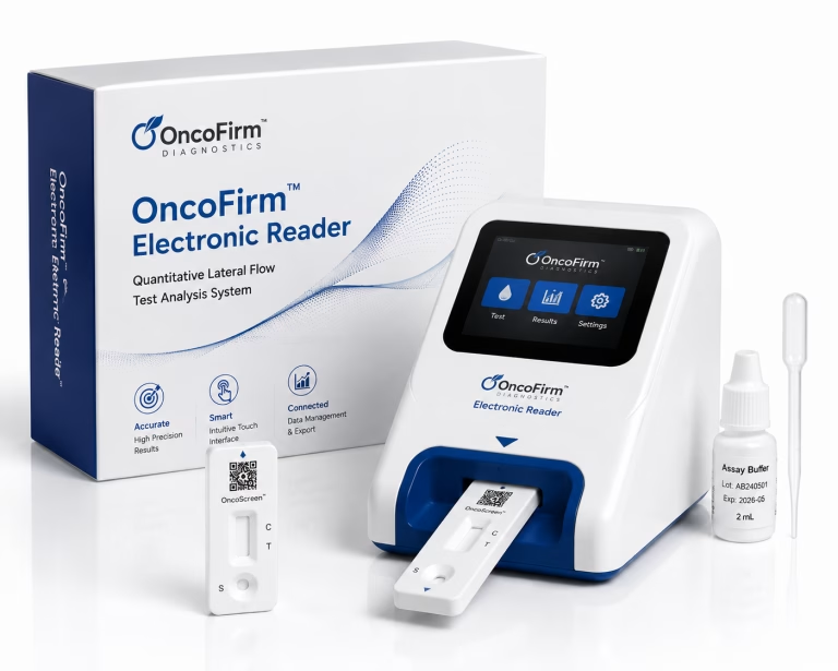
A scientifically defensible OncoFirm development architecture could combine a multiplex fluorescent lateral-flow assay, an electronic reader, analytical quality controls, and a locked multivariable risk algorithm.
This description is a conceptual development framework. It does not represent evidence that an OncoFirm test has established analytical sensitivity, clinical sensitivity, specificity, predictive value, cancer-site localization, or clinical utility.
No numerical clinical-performance claims are assigned in this paper because verified OncoFirm clinical datasets were not provided for review.
12.2 Potential system architecture
A proposed investigational workflow could include:
- Collection of the validated blood specimen type;
- Application of the specimen to a multiplex assay cartridge;
- Detection of selected protein, antigen, antibody, or related biomarker signals;
- Acquisition of raw fluorescence and internal-control signals by an electronic reader;
- Automated cartridge and analytical quality assessment;
- Normalization of biomarker signals;
- Integration with a limited set of clinically justified patient variables;
- Generation of a calibrated risk estimate;
- Assignment to a lower-risk, elevated-risk, indeterminate, or invalid category;
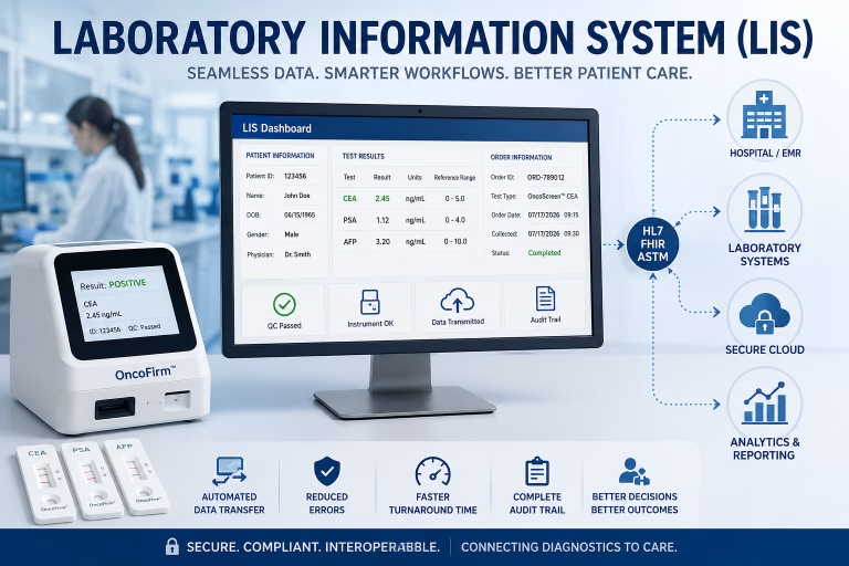
- Secure reporting through the reader, laboratory information system, or electronic medical record;
- Recording of model version, reader, lot, operator, and quality-control data.
12.3 Intended-use-first development
The platform should initially pursue a narrow, clinically defined intended use rather than a broad claim of universal cancer detection.
Potential investigational use cases should be evaluated separately and could include:
- Triage of a specified symptomatic referral population;
- Adjunctive risk stratification following a defined abnormal laboratory result;
- Assessment of a defined high-risk population;
- Support for referral prioritization in a specified clinical pathway;
- Longitudinal monitoring of biomarker patterns, provided the monitoring claim is independently validated.
A population-wide multi-cancer screening claim would require substantially broader evidence, including prospective evaluation of diagnostic pathways, false-positive burden, interval cancers, stage-specific performance, and clinical utility.
12.4 Proposed staged development pathway
| Stage | Principal objective | Required output |
|---|---|---|
| 0. Target product profile | Define population, intended use, specimen, setting, output, and comparator | Approved clinical and regulatory development specification |
| 1. Analytical feasibility | Establish measurable signals and preliminary assay behavior | Candidate biomarkers and stable assay configuration |
| 2. Biomarker and model discovery | Evaluate complementary value in retrospective specimens | Candidate locked panel and provisional model |
| 3. Analytical validation | Characterize precision, sensitivity, interference, stability, reader, and system performance | Validated analytical operating range |
| 4. Independent clinical validation | Test the locked system in external specimens | Prespecified estimates of clinical performance and calibration |
| 5. Prospective silent-mode evaluation | Run the system without influencing care | Real-world workflow, failure, drift, and calibration evidence |
| 6. Prospective interventional evaluation | Measure the consequences of using the result | Evidence of safety and clinical utility |
| 7. Regulatory review and controlled deployment | Establish compliant manufacturing, software, labeling, and surveillance | Authorized intended use and postmarket plan |
12.5 Biomarker-panel selection
Biomarkers should not be selected only because they differ statistically between cancer cases and healthy controls. Selection should consider:
- Independent incremental value;
- Biological plausibility;
- Stability in the intended specimen;
- Analytical compatibility;
- Cross-reactivity;
- benign-condition distributions;
- stage-specific behavior;
- prevalence in the intended population;
- manufacturing feasibility;
- assay dynamic range;
- contribution to calibration and net clinical benefit.
Removal of a biomarker from a model should be evaluated with the same rigor as its addition.
12.6 Reader and software design
The reader should preserve raw and processed data necessary for traceability. Software should distinguish analytical quality-control decisions from clinical risk classification.
A recommended architecture would keep separate modules for:
- Cartridge identification;
- image or optical acquisition;
- signal extraction;
- analytical validity;
- normalization;
- biomarker quantification;
- risk prediction;
- report generation;
- data transmission;
- audit logging.
This modular design can support testing, failure analysis, cybersecurity review, and controlled updates.
12.7 Clinically appropriate claims
Before adequate evidence exists, scientifically appropriate language includes:
- “Investigational”;
- “Under development”;
- “Designed to evaluate”;
- “Intended for prospective validation”;
- “May support risk stratification if clinically validated.”
Language that should not be used without supporting evidence includes:
- “Diagnoses cancer”;
- “Rules out cancer”;
- “Ultra-early detection”;
- “Laboratory-equivalent accuracy”;
- “Higher sensitivity”;
- “Prevents cancer deaths”;
- “Reduces unnecessary procedures”;
- “Detects multiple cancers from a single drop of blood.”
Each of these statements constitutes a testable performance or clinical-utility claim requiring appropriately designed evidence.
13. Research Priorities
Priority areas for the field include:
13.1 Early-stage biological sensitivity
Research should determine which biomarkers are reliably detectable during the preclinical phase and how sensitivity varies by cancer type, subtype, stage, tumor volume, and anatomical site.
13.2 Benign-condition specificity
Development cohorts require sufficient representation of nonmalignant conditions that resemble cancer biologically or clinically.
13.3 Longitudinal measurement
Repeated measurements may distinguish persistent biological change from transient variation. However, longitudinal algorithms require their own analytical, statistical, and clinical validation.
13.4 Clinical-action thresholds
Thresholds should be selected according to the relative harms of false-negative and false-positive results within the intended pathway. They should not be selected solely to maximize a statistical index in the development dataset.
13.5 Diagnostic resolution
Research should define efficient, evidence-based workups after an elevated blood-test result, particularly when the probable cancer origin is uncertain.
13.6 Comparative effectiveness
New tests should be compared with the actual standard diagnostic pathway, not only with no testing. Comparators may include recommended screening, symptom-based referral, routine laboratory assessment, imaging, or established risk models.
13.7 Equity and implementation
Prospective studies should evaluate whether the technology reduces or widens differences in screening participation, diagnostic follow-up, treatment access, and outcomes.
14. Limitations of the Present Review
This paper is a narrative technical review rather than a systematic review or meta-analysis. It does not provide pooled estimates of test performance.
No proprietary OncoFirm assay-development data, reader-performance data, algorithm documentation, clinical datasets, or regulatory submissions were independently reviewed. Consequently, the OncoFirm section describes a proposed scientific and validation framework rather than a verified product-performance assessment.
The field is evolving rapidly. Regulatory requirements, reporting standards, trial outcomes, and professional recommendations should be rechecked immediately before publication or use in development planning.
15. Conclusion
AI-enabled multi-biomarker blood testing offers a plausible method for combining weak, heterogeneous cancer-associated signals into clinically interpretable risk estimates. Its potential arises from the complementarity of molecular, protein, immune, physiological, and patient-level information.
The central translational challenge is not merely whether an algorithm can separate selected cancer cases from selected controls. The more important question is whether the complete test system performs reliably in its intended population and improves a defined clinical pathway without causing disproportionate false-positive investigations, missed cancers, overdiagnosis, anxiety, cost, or inequity.
Population screening and clinical triage must be developed as distinct intended uses. Both require stable assays, representative datasets, locked and calibrated models, external validation, prospective evaluation, human oversight, controlled software governance, and transparent reporting.
For OncoFirm, the scientifically strongest development strategy is an intended-use-first program that integrates assay engineering, reader quality control, AI risk estimation, and prospective clinical validation. Claims should expand only as corresponding analytical and clinical evidence is generated.
Declarations
Ethics approval
Not applicable. This narrative technical review reports no new research involving human participants, human specimens, or identifiable patient information.
Data availability
No new dataset was generated or analyzed for this paper.
Funding
By OncoFirm Diagnostics Corporation
Competing interests
OncoFirm Diagnostics is developing cancer diagnostic technologies and therefore has a commercial interest in the subject matter discussed. This paper does not establish the safety, effectiveness, regulatory status, or clinical utility of any OncoFirm product.
Author contributions
[Identify the human authors responsible for conceptualization, scientific review, regulatory review, writing, and final approval before publication.]
AI-assisted drafting disclosure
This manuscript was drafted with artificial-intelligence-assisted writing support under user direction. Qualified human scientific, medical, statistical, legal, and regulatory reviewers should verify all claims, references, disclosures, and terminology before publication. Final authorship and accountability must remain with named human authors.
Read the full OncoFirm™ Diagnostics whitepaper:
https://oncofirmdiagnostics.com/ai-multi-biomarker-blood-tests-cancer-screening-triage/
Connect with OncoFirm™ Diagnostics
Website: https://oncofirmdiagnostics.com
Whitepapers: https://oncofirmdiagnostics.com/category/whitepapers/
LinkedIn: https://www.linkedin.com/company/oncofirm-diagnostics/
Instagram: https://www.instagram.com/oncofirm/
Facebook: https://www.facebook.com/oncofirm/
Email: [email protected]
Telephone: +1 (516) 900-2606
OncoFirm™ Diagnostics — Advancing earlier cancer detection through biomarker science, fluorescent lateral flow technology, digital diagnostics, and responsible artificial intelligence.
Reference Placeholder Map
Replace each placeholder with verified, properly formatted references before publication.
| Placeholder | Evidence required |
|---|---|
| R1 | Authoritative definition, terminology, current clinical status, benefits, harms, and limitations of blood-based single- or multi-cancer detection |
| R2 | Primary study of a multi-analyte blood test combining circulating DNA and protein biomarkers |
| R3 | Primary validation study of a methylation-based multi-cancer classifier |
| R4 | Primary study of cell-free DNA fragmentation or fragmentomics |
| R5 | Prospective implementation or return-of-results study for multi-cancer blood testing |
| R6 | Primary diagnostic-accuracy study of a machine-learning blood-test model in symptomatic or referred patients |
| R7 | Trial-design or clinical-utility framework for multi-cancer screening |
| R8 | Applicable FDA, CLSI, ISO, or other recognized analytical-validation requirements |
| R9 | STARD-AI reporting guideline |
| R10 | TRIPOD+AI reporting guideline |
| R11 | CONSORT-AI and SPIRIT-AI reporting guidance |
| R12 | FDA or IMDRF Good Machine Learning Practice principles |
| R13 | FDA guidance on predetermined change control plans for AI-enabled device software functions |
| R14 | Current professional recommendations for established cancer screening |
| R15 | Primary or consensus evidence addressing health equity, access, and downstream diagnostic burden |
| R16 | Primary analytical study supporting fluorescent lateral-flow quantification, reader performance, or multiplex measurement |
Suggested Evidence Anchors for Editorial Review
These sources can be used to begin replacing the placeholders; the final bibliography should be checked by a medical editor immediately before publication.
- National Cancer Institute. Questions and Answers about Multi-Cancer Detection Tests. Useful for R1 and R14. The NCI describes MCD tests as predictive rather than diagnostic, identifies relevant biomarker classes, and emphasizes the need for confirmatory evaluation and randomized clinical evidence.
- Clarke CA, et al. Lexicon for blood-based early detection and screening: BLOODPAC consensus document. Clinical and Translational Science. 2024;17:e70016. Useful for R1.
- Cohen JD, et al. Detection and localization of surgically resectable cancers with a multi-analyte blood test. Science. 2018. Useful for R2.
- Klein EA, et al. Clinical validation of a targeted methylation-based multi-cancer early detection test using an independent validation set. Annals of Oncology. 2021;32(9). Useful for R3.
- Cristiano S, et al. Genome-wide cell-free DNA fragmentation in patients with cancer. Nature. 2019;570:385–389. Useful for R4.
- Lennon AM, et al. Feasibility of blood testing combined with PET-CT to screen for cancer and guide intervention. Science. 2020;369:eabb9601. Useful for R5.
- Schrag D, et al. Blood-based tests for multicancer early detection: the PATHFINDER prospective cohort study. The Lancet. 2023;402:1251–1260. Useful for R5.
- Savage R, et al. Development and validation of multivariable machine-learning algorithms to predict risk of cancer in symptomatic patients referred urgently from primary care. BMJ Open. 2022;12:e053590. Useful for R6.
- Minasian LM, et al. Study design considerations for trials to evaluate multicancer early detection assays for clinical utility. Journal of the National Cancer Institute. 2023;115:250–257. Useful for R7.
- Sounderajah V, et al. The STARD-AI reporting guideline for diagnostic accuracy studies using artificial intelligence. Nature Medicine. 2025;31:3283–3289. Useful for R9.
- Collins GS, et al. TRIPOD+AI statement: updated guidance for reporting clinical prediction models that use regression or machine-learning methods. BMJ. 2024;385:e078378. Useful for R10.
- Liu X, et al.; Rivera SC, et al. CONSORT-AI and SPIRIT-AI extensions. Useful for R11.
- U.S. Food and Drug Administration and International Medical Device Regulators Forum. Good Machine Learning Practice for Medical Device Development. Useful for R12.
- U.S. Food and Drug Administration. Marketing Submission Recommendations for a Predetermined Change Control Plan for Artificial Intelligence-Enabled Device Software Functions. Useful for R13.