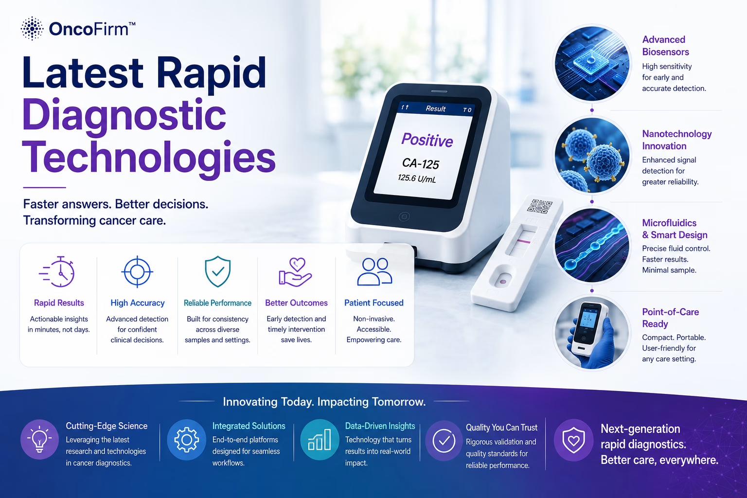
Rapid cancer diagnostic technologies: a map of methods and maturity
By OncoFirm Diagnostics · Last updated October 2026
Rapid cancer diagnostic technologies are assay formats and instruments designed to measure a cancer-associated biomarker near the point of sampling, without sending the specimen to a central laboratory. The main families are lateral flow immunoassays, microfluidic cartridges, biosensors, multiplexed panels and the digital readers that quantify them, and they differ widely in maturity.
Several are well established for other uses and still at research stage for cancer biomarkers. This overview maps the families and points to longer articles on each; it is not a news digest.
The technology landscape at a glance
| Technology | How it works | Maturity | Main open question for oncology |
|---|---|---|---|
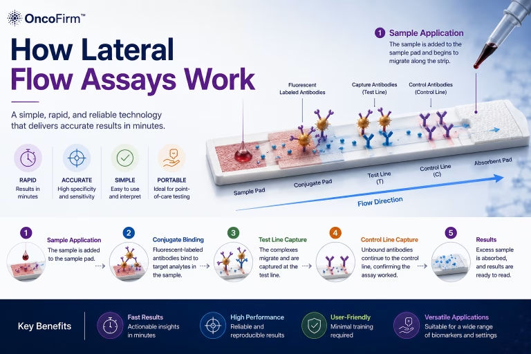
| Colorimetric lateral flow | Labeled antibodies capture the analyte on a strip; a colored line is read by eye | Mature; mass-manufactured for pregnancy and infectious-disease testing | Whether a visual readout suits biomarkers that need quantification |
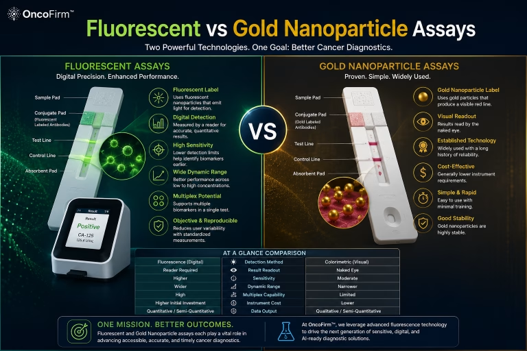
| Fluorescent lateral flow with a reader | Same strip architecture with fluorescent labels; a reader measures line intensity | Commercial for some analytes; research stage for most cancer biomarkers | Reader-to-reader and lot-to-lot reproducibility |
| Microfluidic cartridges | Channels move metered sample and reagents through timed steps | Used in some point-of-care analyzers; many oncology designs are prototypes | Cost and robustness outside the laboratory |
| Biosensors | A recognition element is coupled to an electrical or optical transducer | Glucose meters are established; cancer sensors are largely proof of concept | Matrix effects in blood; sensor stability |
| Multiplexed panels | Several analytes are measured in one run | Routine on laboratory platforms; harder in rapid formats | Cross-reactivity; how results are combined |
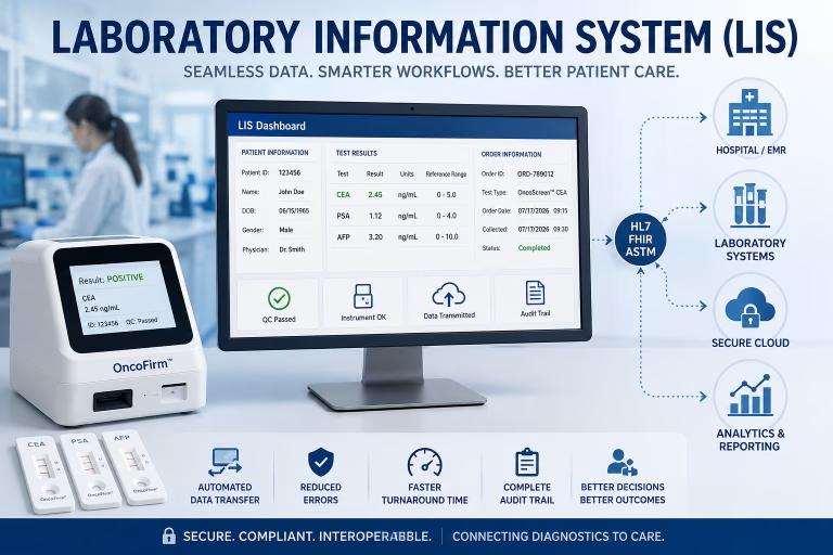
| Digital readers and software | Optics and algorithms convert a signal into a number | Established for fluorescent strips; smartphone readers are mostly research tools | Standardization and software validation |
Maturity describes each format in general, not a specific product.
Lateral flow: the most mature rapid format
Lateral flow assays are inexpensive to make, need small sample volumes and often do not require cold storage, which explains their reach (Posthuma-Trumpie et al., 2009). Their recognized weaknesses are limited sensitivity with visual labels, restricted quantification and variation between lots.
Colloidal gold, colored latex, fluorescent dyes, quantum dots and upconverting particles each bring different signal and reader requirements (Bahadır and Sezgintürk, 2016). The trade-offs are set out in our comparison of fluorescent and gold nanoparticle labels, and the strip mechanics in how lateral flow assays work.
Microfluidics and biosensors: more control, more complexity
Microfluidic cartridges add what a simple strip lacks: metered volumes, timed incubation, wash steps and sometimes plasma separation. Those steps can improve precision, but each adds parts, cost and failure modes.
Biosensors replace the optical line with a transducer. Nanomaterials have been used to raise signal in both strip-based and sensor-based designs (Quesada-González and Merkoçi, 2015). Many published cancer biomarker sensors have been characterized in buffer or in small specimen sets; performance in clinical specimens across sites is the evidence most often missing.
Multiplexing and digital readout
Most serum tumor markers lack the sensitivity and specificity to detect early cancer when used alone (Duffy, 2013), which is why panels attract interest. Putting several antibody pairs on one strip raises problems that single-analyte tests avoid: cross-reactivity, different optimal conditions for each analyte, and the need for a prespecified rule for combining results.
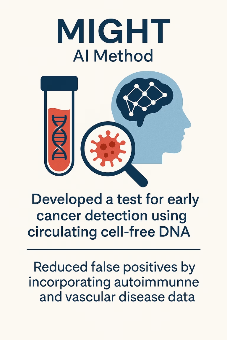
A digital reader turns line intensity into a number and can apply quality-control checks before reporting. OncoFirm™ is developing a prototype portable digital fluorescence reader for its research-stage assay platform; its performance has not been established. Software that interprets the signal raises separate validation questions, covered in AI in cancer diagnostics.
How to judge maturity in any rapid format
A published detection limit is only the start. Four further questions separate a prototype from a usable test.
- Analytical validation. Precision, detection limit, interference and stability in the intended specimen type.
- Clinical validation. Performance in the intended-use population against an accepted reference standard, including people with benign conditions.
- Manufacturability. Whether lots can be made reproducibly at scale under a quality system.
- Use in context. Whether non-specialist operators obtain the same result, and whether the result changes a clinical decision (Drain et al., 2014).
Speed alone does not make a test useful. What a rapid result would be used for is taken up in the future of cancer screening.
Research gaps where partners can contribute
The open questions suit specific partners: antibody and reagent developers for pair selection and cross-reactivity, optical and microfluidic engineers for reader reproducibility, clinical laboratories for method comparison against reference immunoassays, and biobanks for well-characterized specimens. OncoFirm is seeking such partners for joint grant applications.
Current questions are listed under research and development priorities, and proposals can be raised through the collaboration opportunities page.
Frequently asked questions
Are rapid tests used in cancer care today?
A few immunoassay-based tests are established, most visibly fecal immunochemical testing in colorectal cancer screening. For most blood-based cancer biomarkers, rapid formats remain research tools.
Is a fluorescent lateral flow assay more sensitive than a gold-based one?
It can be in some assay designs, because a reader can measure signals too faint to see. The gain has to be demonstrated for each assay.
Does OncoFirm sell a rapid cancer test?
No. OncoFirm’s assay, reader and software are in research and development, with no established performance and no regulatory clearance.
Related research and resources
- Tumor antigen detection — the targets that rapid immunoassays try to measure.
- Point-of-care oncology — where near-patient testing fits.
- Funding and research collaboration opportunities — how laboratories and companies can join.
References and further reading
- Posthuma-Trumpie GA, Korf J, van Amerongen A. Lateral flow (immuno)assay: its strengths, weaknesses, opportunities and threats. A literature survey. Anal Bioanal Chem. 2009;393(2):569–582.
- Bahadır EB, Sezgintürk MK. Lateral flow assays: principles, designs and labels. TrAC Trends Anal Chem. 2016;82:286–306.
- Quesada-González D, Merkoçi A. Nanoparticle-based lateral flow biosensors. Biosens Bioelectron. 2015;73:47–63.
- Duffy MJ. Tumor markers in clinical practice: a review focusing on common solid cancers. Med Princ Pract. 2013;22(1):4–11.
- Drain PK, Hyle EP, Noubary F, et al. Diagnostic point-of-care tests in resource-limited settings. Lancet Infect Dis. 2014;14(3):239–249.
Research and regulatory status
OncoFirm™ technologies described on this site are in research and development. Their performance characteristics have not been established, and no OncoFirm product has been cleared, approved or authorized by the U.S. Food and Drug Administration. Nothing here is medical advice. Reference to a federal agency or funding program does not imply endorsement, funding, sponsorship or affiliation.