Tumor antigen detection: antigen classes, assays and interpretation
By OncoFirm Diagnostics · Last updated October 2026
Tumor antigen detection is the measurement, with antibodies, of proteins or carbohydrate structures that tumors produce or shed into blood and other fluids. In diagnostics it almost always means tumor-associated antigens such as CEA, CA-125 or PSA, which normal tissue also makes, and that is the root of its main limitation: specificity.
Immunologists use the term differently, for whatever the immune system can recognize on a cancer cell. The two usages overlap without being identical, and the difference decides which antigens can realistically be measured in a blood sample.
Tumor-associated antigens, tumor-specific antigens and neoantigens
| Class | Origin | Examples | Relevance to blood-based detection |
|---|---|---|---|
| Tumor-associated antigens | Normal proteins or glycans that are overexpressed, re-expressed from fetal development or restricted to one tissue | CEA, AFP, CA-125, CA 19-9, PSA | The basis of most serum tumor marker assays; also raised in some benign conditions |
| Tumor-specific antigens | Molecules absent from normal cells, including viral oncoproteins and mutant proteins | HPV E6 and E7; products of tumor mutations | Studied mainly as immunotherapy targets, not as serum markers |
| Neoantigens | The subset of tumor-specific antigens created by somatic mutation, often unique to one patient’s tumor | Mutant peptides presented on MHC molecules | Identified by sequencing and prediction, not by a standard immunoassay |
Tumor-associated antigens dominate diagnostics because they are released in measurable quantities. How antigens relate to DNA, RNA and cellular markers is set out in what are cancer biomarkers.
Immunoassay formats used to measure antigens
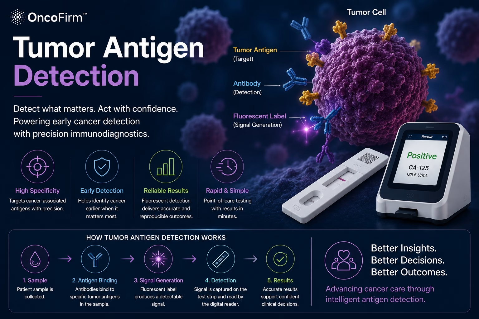
Most antigen assays are sandwich immunoassays: one antibody captures the target and a second, labeled antibody reports it. The formats differ in where that happens. ELISA uses a plate with wash steps; automated chemiluminescent analyzers do the same at scale in hospital laboratories; bead-based systems measure many analytes at once for research. Strip formats put the sandwich on a membrane, as explained in how lateral flow assays work, with label options compared in fluorescent vs gold nanoparticle lateral flow assays.
Results are not interchangeable between methods. Assays from different manufacturers for one marker use antibodies to different sites and are not fully standardized, which is why serial monitoring should stay on one method.
Why a single marker has limited specificity
Most serum markers rise in conditions other than cancer: CA-125 with endometriosis and peritoneal inflammation, CEA in smokers and in inflammatory bowel disease, PSA with benign prostatic enlargement, AFP with hepatitis and cirrhosis. PSA is specific to an organ, not to a tumor.
Sensitivity is limited from the other side. Small tumors shed little antigen, not every tumor of a type expresses the marker, and people who lack the Lewis blood group antigen produce little or no CA 19-9. Together these explain why established markers are used mainly for monitoring and surveillance, with few suited to screening (Duffy, 2013).
How marker panels are being studied
Because tumors of one type differ in what they express (Duffy, 2013), combining markers can raise sensitivity. The catch is that each added marker also adds its own false positives unless the results are combined in a prespecified model whose overall specificity has been validated. Algorithms that pair CA-125 with HE4 for women with a pelvic mass are an example already in clinical use; research panels have gone further and combined proteins with mutations in circulating DNA (Cohen et al., 2018).
A panel is a prediction model, and it fails in the ways models fail: overfitting to the discovery set and optimistic performance that shrinks on independent data. Reporting guidance such as TRIPOD+AI sets out what a credible evaluation must disclose (Collins et al., 2024). Our research framework summary on multi-biomarker blood tests takes the topic further.
Reading antigen results alongside imaging and pathology
An antigen concentration is one input. Diagnosis of a solid tumor rests on imaging and tissue pathology; an abnormal marker changes the probability of disease and can prompt that workup, but it does not replace it.
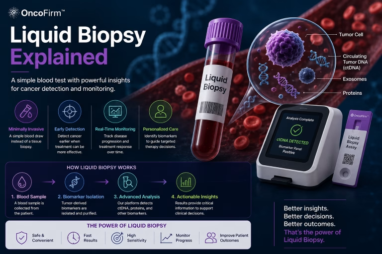
In monitoring, the trend matters more than any single value: a sustained rise on the same assay is a reason to image, not proof of recurrence. Protein measurements also say nothing about tumor genotype, which is the territory of the DNA-based methods in liquid biopsy explained.
Where antibody and cohort partners fit
OncoFirm is developing a research-stage fluorescent lateral flow immunoassay platform for cancer-associated protein biomarkers; the approach is outlined on its antigen technology page. Which antigens and combinations merit a place on the platform is an open research question, and no performance has been established.
Progress depends on antibody developers with well-characterized reagent pairs, on groups holding serum sets that include benign disease controls as well as cancers, and on biostatisticians to prespecify how markers are combined. To propose a joint study on marker selection or validation, use the form to discuss a grant opportunity.
Frequently asked questions
What is the difference between a tumor-associated and a tumor-specific antigen?
A tumor-associated antigen is a normal molecule that tumors make in larger amounts or in the wrong context, so it is also found in healthy tissue. A tumor-specific antigen, such as a mutant protein or a viral oncoprotein, is absent from normal cells.
Is a raised tumor marker proof of cancer?
No. Most markers rise in benign conditions as well, and some cancers do not raise them. A raised value is a reason for further investigation, interpreted with imaging and pathology.
Why do results for the same marker differ between laboratories?
Assays from different manufacturers use different antibodies and calibrators, so values are not directly comparable. Serial measurements in one patient should use the same method.
Are neoantigens measured in blood tests?
Not routinely. Neoantigens are short mutant peptides displayed on tumor cells and are identified by sequencing the tumor, mainly to guide immunotherapy research.
Related research and resources
- What are cancer biomarkers? — where protein antigens sit among the other biomarker classes
- Liquid biopsy explained — circulating DNA and cells, and how they complement protein markers
- How lateral flow assays work — the strip format for near-patient antigen measurement
- Clinical collaborations — how groups holding clinical specimens can take part
References and further reading
- Duffy MJ. Tumor markers in clinical practice: a review focusing on common solid cancers. Med Princ Pract. 2013;22(1):4–11.
- National Cancer Institute. Tumor Markers — fact sheet.
- Cohen JD, Li L, Wang Y, et al. Detection and localization of surgically resectable cancers with a multi-analyte blood test. Science. 2018;359(6378):926–930.
- Collins GS, Moons KGM, Dhiman P, et al. TRIPOD+AI statement: updated guidance for reporting clinical prediction models that use regression or machine learning methods. BMJ. 2024;385:e078378.
Research and regulatory status
OncoFirm™ technologies described on this site are in research and development. Their performance characteristics have not been established, and no OncoFirm product has been cleared, approved or authorized by the U.S. Food and Drug Administration. Nothing here is medical advice. Reference to a federal agency or funding program does not imply endorsement, funding, sponsorship or affiliation.