Fluorescent vs gold nanoparticle lateral flow assays compared
By OncoFirm Diagnostics · Last updated October 2026
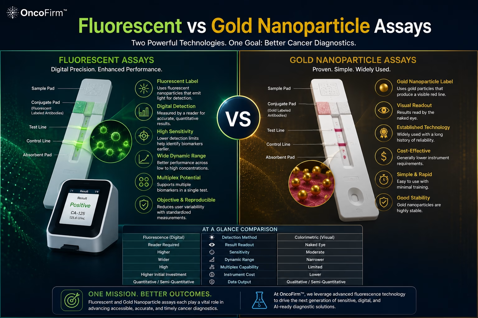
Fluorescent and gold nanoparticle lateral flow assays share the same strip and the same antibody chemistry; they differ in the label attached to the detection antibody and in how the signal is read. Gold gives a colored line visible to the eye, while fluorescent labels emit light that an instrument measures, which often lowers the detection limit and yields a number at the price of needing a reader.
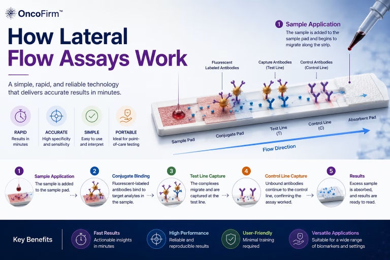
Neither is better in general. The choice depends on how low the analyte concentration is, whether a number or a yes-or-no answer is required, and where the test will be run. The strip mechanics common to both are covered in how lateral flow assays work.
How each label produces a signal
Colloidal gold particles, typically tens of nanometers across, absorb green light strongly and so appear red. When enough of them collect at the test line, a line becomes visible. Antibodies attach readily to the gold surface, and gold has long been the most widely used label (Posthuma-Trumpie et al., 2009; Sajid et al., 2015).
Fluorescent labels include dye-loaded polymer microspheres, europium chelate particles, quantum dots and upconverting phosphors (Bahadır and Sezgintürk, 2016; Quesada-González and Merkoçi, 2015). A reader excites the label and measures the emitted light. Some labels are chosen to suppress background: europium chelates emit long after excitation, so short-lived background fluorescence can be gated out, and upconverting particles emit at a shorter wavelength than the near-infrared light that excites them, which background fluorescence cannot do.
Side-by-side comparison
| Property | Gold nanoparticle label | Fluorescent label |
|---|---|---|
| Signal | Light absorption; a colored line | Light emission after excitation |
| Readout | Eye, or a reflectance reader or camera | Dedicated fluorescence reader |
| Result type | Qualitative by eye; semi-quantitative with a reader | Quantitative or semi-quantitative, if calibrated |
| Detection limit | Suited to abundant analytes; can be extended by signal enhancement | Often lower in published comparisons; limited by background and antibody affinity |
| Main error sources | Subjective reading of faint lines; lighting | Membrane and sample autofluorescence; photobleaching of some dyes; reader drift |
| Multiplexing | Separate lines or strips | Separate lines, or spectrally distinct labels |
| Cost and logistics | Lowest; no instrument needed | Higher; instrument, calibration and maintenance |
| Track record | Long, across many analytes | Shorter; commercial systems exist for analytes such as cardiac and inflammatory markers |
General properties of the two label types as described in the review literature. Performance is specific to each assay and has to be measured.
Why a lower detection limit is not automatically a better test
Label brightness is one term in the detection limit. Antibody affinity, non-specific binding and background set the floor just as firmly, and a brighter label amplifies non-specific signal along with specific signal.
For proteins that healthy people also carry, the clinical question is rarely whether any is present. It is whether the concentration sits above or below a cutoff, so precision near that cutoff matters more than the lowest detectable amount. The reasons are set out in tumor antigen detection.
Strip tests against central laboratory immunoassay
Strip formats are easier to place when set beside the method they are usually compared with. These are general properties of each format, not performance claims for any product.
Central laboratory immunoassay
Automated analyzers, commonly chemiluminescent, run by trained staff with wash steps, multi-point calibration, internal quality control and external quality assessment. Quantitative and high-throughput, but dependent on a laboratory and sample transport.
Conventional visual lateral flow
A single-use strip read by eye. It needs no instrument and little training, so it reaches settings a laboratory cannot. Qualitative, with variation between observers on faint lines and no automatic record.
Reader-based fluorescent lateral flow
The same strip with an instrument readout. It gives a numerical, recordable result near the patient, but depends on lot calibration and reader quality control, and still lacks an analyzer’s wash steps.
Choosing between the two labels
Gold remains the sensible choice when the analyte is abundant, a binary answer is enough and no instrument can be supported. Fluorescence earns its extra cost when concentrations are low, when a value must be compared with a cutoff or followed over time, and when the setting can maintain a reader.
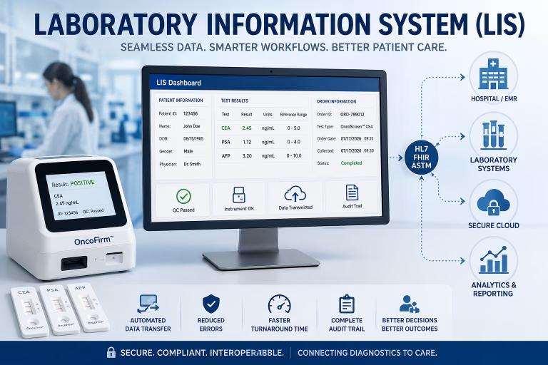
A reader also produces a digital record, which is what allows software interpretation and transfer to clinical systems. Those requirements are discussed in point-of-care oncology testing and, on the software side, under AI-assisted signal analysis.
Label and reader questions open to collaborators
OncoFirm™ is developing a fluorescent lateral flow immunoassay platform and a prototype portable digital fluorescence reader. Both are research-stage, and no performance has been established. Open questions include label selection and conjugate stability, background reduction on the membrane, agreement between reader units, and method comparison against laboratory immunoassays.
Nanoparticle and reagent suppliers, optical engineers and clinical laboratories are the partners this work needs. Start with the research collaboration inquiry.
Frequently asked questions
Are fluorescent lateral flow assays always more sensitive than gold?
No. Fluorescent labels often lower the detection limit, but the result depends on antibody quality, background and reader design. A well-optimized gold assay can outperform a poorly optimized fluorescent one.
Why is colloidal gold still so widely used?
It is inexpensive, stable, simple to conjugate to antibodies and visible without an instrument. For abundant analytes and yes-or-no questions those advantages are hard to beat.
Does a fluorescent lateral flow assay need a reader?
Yes. The label has to be excited at a suitable wavelength and its emission measured by a detector, and any quantitative result also depends on the reader applying a calibration.
Can a gold nanoparticle strip be read by an instrument?
Yes. Reflectance readers and cameras can measure colored line intensity and give semi-quantitative results, which narrows but does not remove the difference from fluorescent systems.
Related research and resources
- How lateral flow assays work — strip anatomy, assay formats and failure modes common to both labels
- Point-of-care oncology testing — what must be shown before any near-patient oncology test is used
- Digital fluorescence reader — the prototype reader OncoFirm is developing
- Laboratory information systems — where reader results have to go to be clinically usable
References and further reading
- Posthuma-Trumpie GA, Korf J, van Amerongen A. Lateral flow (immuno)assay: its strengths, weaknesses, opportunities and threats. A literature survey. Anal Bioanal Chem. 2009;393(2):569–582.
- Bahadır EB, Sezgintürk MK. Lateral flow assays: principles, designs and labels. TrAC Trends Anal Chem. 2016;82:286–306.
- Quesada-González D, Merkoçi A. Nanoparticle-based lateral flow biosensors. Biosens Bioelectron. 2015;73:47–63.
- Sajid M, Kawde AN, Daud M. Designs, formats and applications of lateral flow assay: a literature review. J Saudi Chem Soc. 2015;19(6):689–705.
Research and regulatory status
OncoFirm™ technologies described on this site are in research and development. Their performance characteristics have not been established, and no OncoFirm product has been cleared, approved or authorized by the U.S. Food and Drug Administration. Nothing here is medical advice. Reference to a federal agency or funding program does not imply endorsement, funding, sponsorship or affiliation.