AI in cancer diagnostics: uses, AI-driven biomarkers and validation
By OncoFirm Diagnostics · Last updated October 2026
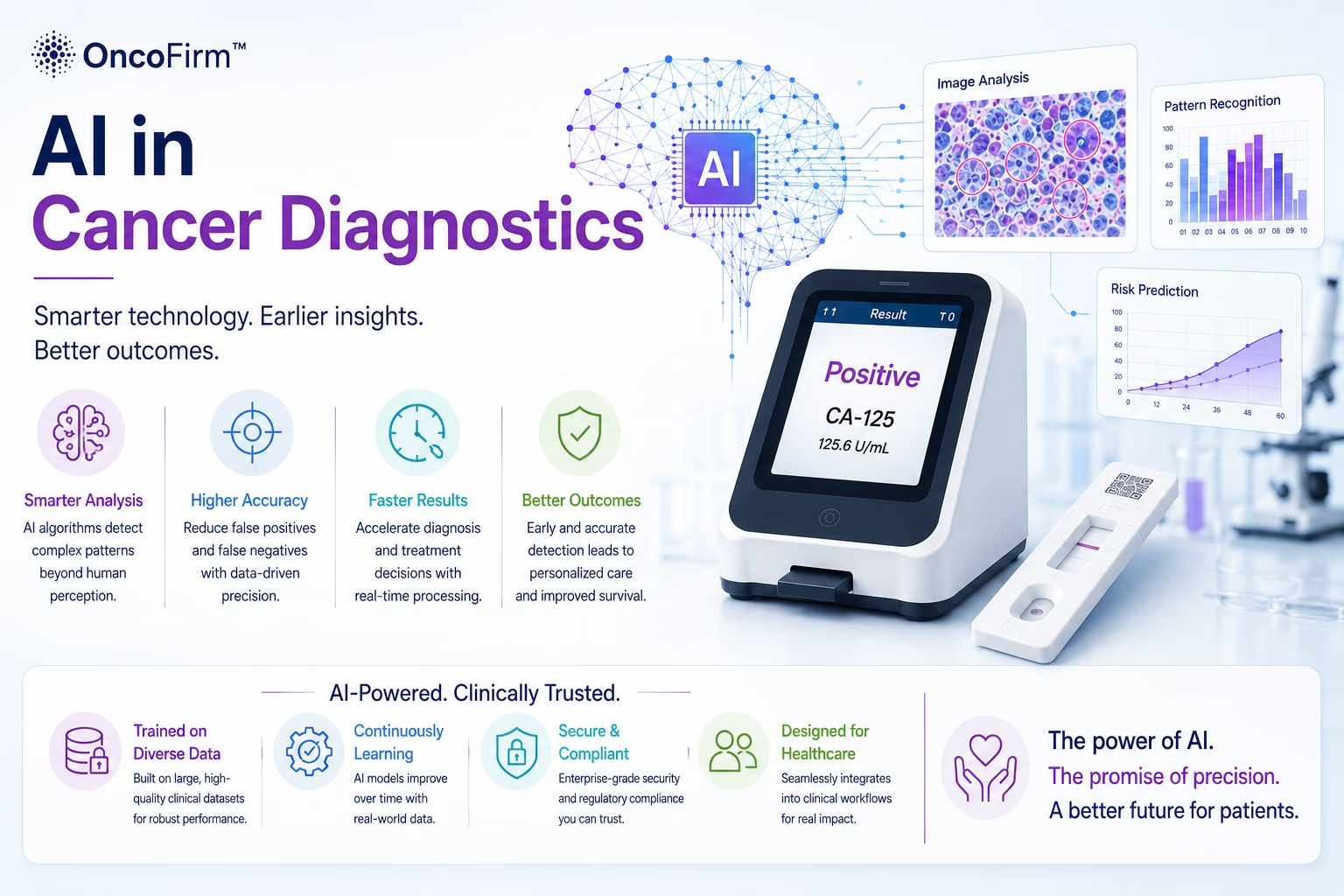
AI in cancer diagnostics means using machine-learning models to interpret images, assay signals, molecular data or clinical records in support of a decision made by a clinician or laboratory professional. The evidence is most developed for image-based tasks in radiology and digital pathology (Topol, 2019; Bera et al., 2019); for blood-based biomarker interpretation it is at an earlier stage, and prospective validation is the main gap.
Below: where models are applied, what “AI-driven biomarker” means, and the methodological problems that most often undermine diagnostic AI studies.
Where AI is used along the diagnostic pathway
Four application areas recur in the literature.
Medical imaging
Models flag or segment suspicious regions in mammography, CT and MRI.
Digital pathology
Models classify tissue, count cells and support grading on whole-slide images.
Assay and biomarker signals
Algorithms normalize raw signals, check quality controls and combine several analytes into one score.
Clinical records and risk models
Prediction models use history, routine laboratory results and demographics to estimate risk.
AI-driven biomarkers: what the term means
An AI-driven biomarker is a measurable indicator whose value is computed by a trained model from many input features, instead of being read from a single analyte. The inputs may be DNA fragment profiles, pixel patterns in a scan or several protein concentrations; the output is a score or a class.
One published example is the work of Cristiano et al. (2019), who applied machine learning to genome-wide cell-free DNA fragmentation profiles. It was a case-control study, so it shows that a signal exists, not how a test would perform in screening.
Multimodal models combine data types:
- Genomic and epigenomic data: mutations, methylation and fragmentation patterns.
- Imaging: quantitative features from CT, MRI or mammography.
- Histopathology: features from digitized tissue slides.
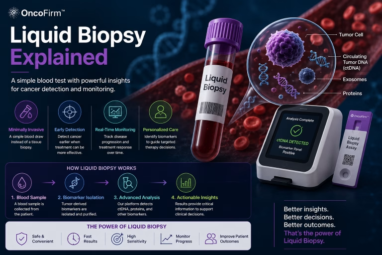
- Liquid biopsy: circulating DNA, proteins and other analytes in blood, described in our liquid biopsy explainer.
- Clinical records: age, history, symptoms and routine laboratory values.
A computed score is still a biomarker. It needs the same analytical and clinical validation as any other, as set out in what cancer biomarkers are, and the model version becomes part of the test.
Validation pitfalls that undermine diagnostic AI
The recurring problems lie in how data are assembled and evaluated.
- Dataset shift. A model trained at one site, on one instrument or in one population often performs worse on data from elsewhere.
- Leakage. Information unavailable at the time of testing, or samples from the same patient, appear in both training and test sets and inflate results.
- Spectrum bias. Comparing advanced cancers with healthy volunteers makes discrimination look easier than it is in clinical use.
- Subgroup bias. Performance differs by age, sex, ancestry or comorbidity when groups are under-represented.
- Models that keep changing. Evaluation needs a locked model and prespecified thresholds; a model altered after test data have been seen has no defined performance.
TRIPOD+AI sets out what a prediction-model study should report (Collins et al., 2024), and CONSORT-AI extends trial reporting to interventions with an AI component (Liu et al., 2020). The Good Machine Learning Practice guiding principles issued by the FDA, Health Canada and the MHRA (2021) add expectations on independent test sets, representative data, the performance of the clinician and software together, and monitoring after deployment.
AI at the point of care: interpreting assay signals
For a reader-based immunoassay, the near-term role of software is specific: locate test and control lines, subtract background, reject invalid runs and convert intensity into a calibrated value. Combining analytes into a risk score is a later, separately validated step. The hardware is covered in our map of rapid cancer diagnostic technologies.
OncoFirm™ is developing AI-assisted signal interpretation software for its research-stage fluorescent lateral flow platform and prototype reader. Its performance has not been established. A staged evaluation plan is summarized in our framework for AI multi-biomarker blood tests.
Where data scientists and biostatisticians come in
OncoFirm’s planned grant projects raise questions that need outside expertise: how to build training and test sets that are independent at patient and site level, how to quantify reader-to-reader variation before any model is trained, and how to prespecify an analysis plan consistent with TRIPOD+AI. Machine-learning groups, biostatistics cores and clinical sites able to contribute annotated specimens are the partners sought.
See the research and development priorities and the collaboration opportunities page.
Frequently asked questions
What is an AI-driven biomarker?
A score or class computed by a trained model from many measurements, such as DNA fragmentation patterns or image features, and used as an indicator of a biological state.
Can AI detect cancer from a blood test?
In research studies, models can separate blood samples from people with and without cancer. Whether a given test works in people without symptoms has to be shown prospectively.
Has OncoFirm validated an AI model?
No. OncoFirm’s signal interpretation software is in development and has no established performance.
Related research and resources
- The future of cancer screening — why test-set accuracy is not clinical benefit.
- Laboratory information system integration — why the model version travels with each result.
- Clinical collaborations — how clinical sites can take part.
References and further reading
- Topol EJ. High-performance medicine: the convergence of human and artificial intelligence. Nat Med. 2019;25(1):44–56.
- Bera K, Schalper KA, Rimm DL, Velcheti V, Madabhushi A. Artificial intelligence in digital pathology — new tools for diagnosis and precision oncology. Nat Rev Clin Oncol. 2019;16(11):703–715.
- Cristiano S, Leal A, Phallen J, et al. Genome-wide cell-free DNA fragmentation in patients with cancer. Nature. 2019;570(7761):385–389.
- Collins GS, Moons KGM, Dhiman P, et al. TRIPOD+AI statement: updated guidance for reporting clinical prediction models that use regression or machine learning methods. BMJ. 2024;385:e078378.
- Liu X, Cruz Rivera S, Moher D, Calvert MJ, Denniston AK; SPIRIT-AI and CONSORT-AI Working Group. Reporting guidelines for clinical trial reports for interventions involving artificial intelligence: the CONSORT-AI extension. Nat Med. 2020;26(9):1364–1374.
- U.S. Food and Drug Administration, Health Canada, MHRA. Good Machine Learning Practice for Medical Device Development: Guiding Principles. 2021.
Research and regulatory status
OncoFirm™ technologies described on this site are in research and development. Their performance characteristics have not been established, and no OncoFirm product has been cleared, approved or authorized by the U.S. Food and Drug Administration. Nothing here is medical advice. Reference to a federal agency or funding program does not imply endorsement, funding, sponsorship or affiliation.