How lateral flow assays work: strip anatomy, formats and limits
By OncoFirm Diagnostics · Last updated October 2026
A lateral flow assay works by letting a liquid sample wick along a strip of porous membrane, carrying labeled antibodies past a line of immobilized capture reagent. If the target is present, label accumulates at that line and produces a signal that is read by eye or by an instrument.
The format is familiar from pregnancy tests and infectious disease rapid tests, and its weaknesses are as well documented as its strengths (Posthuma-Trumpie et al., 2009). Both matter for cancer-associated protein biomarkers, where a quantitative result near a cutoff is usually needed and a yes-or-no answer is not enough.
Anatomy of a test strip
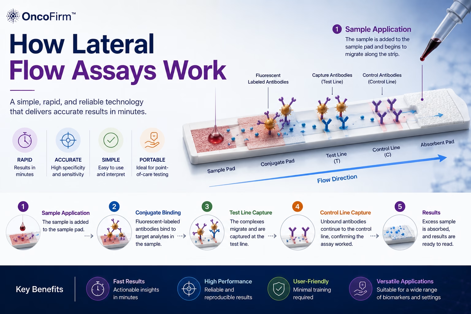
Four overlapping porous materials are laminated onto a backing card, and each has a job.
| Component | Function | Typical failure point |
|---|---|---|
| Sample pad | Receives the specimen; filters, buffers and meters it onto the strip | Viscous or cellular samples flow unevenly |
| Conjugate pad | Holds dried, labeled detection antibody that redissolves into the sample | Incomplete or variable release of the conjugate |
| Nitrocellulose membrane | Carries the test and control lines of immobilized capture reagent | Capillary flow rate differs between membrane lots |
| Absorbent pad | Wicks liquid through the strip and sets the volume drawn | Saturation allows backflow |
| Backing and cassette | Hold the parts in contact; fix the sample port and read window | Poor overlap or compression interrupts flow |
Several analytes can be measured on one strip by adding test lines.
From sample to signal, step by step
- The sample (whole blood, plasma, serum, urine or saliva, often with a running buffer) is applied to the sample pad.
- Capillary action draws it into the conjugate pad, where it rehydrates the labeled antibody. Any target present binds the label.
- The mixture migrates along the membrane. At the test line, immobilized capture antibody binds the target-label complex and concentrates the label.
- Further along, the control line captures surplus labeled antibody whether or not target is present, confirming that liquid flowed and reagents were active.
- After a fixed development time the lines are read. Timing matters, because the signal keeps changing as the strip dries.
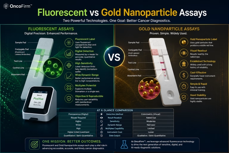
No pump or power is involved; flow is set by the membrane’s pore structure and the sample’s viscosity. The label itself, colored or fluorescent, is compared in fluorescent vs gold nanoparticle lateral flow assays.
Sandwich and competitive formats
In the sandwich format, two antibodies bind different sites on the same target: one carries the label, the other is fixed at the test line. Signal rises with concentration. Proteins are large enough to offer two binding sites, so most assays for the markers described in tumor antigen detection are sandwiches.
The competitive format is used for small molecules that cannot bind two antibodies at once. Target in the sample competes with target fixed at the test line for a limited amount of labeled antibody, so signal falls as concentration rises (Sajid et al., 2015).
Where the format struggles
- Hook effect. In a one-step sandwich, a very high target concentration saturates the labeled and capture antibodies separately, so fewer sandwiches form and a high sample reads falsely low. Tumor marker concentrations can span several orders of magnitude, which makes this a practical concern.
- Matrix effects. Hematocrit, viscosity and lipids change flow. Interfering antibodies in some patients’ blood can bridge capture and detection antibodies without any target.
- Lot variability. Membranes, conjugates and dispensed lines vary between batches, and a single-use strip cannot be calibrated individually, so calibration is assigned per lot.
- No wash step. Unbound label is not removed as it is in a laboratory immunoassay, which raises background and limits analytical sensitivity.
Each of these is described in the review literature (Posthuma-Trumpie et al., 2009; Bahadır and Sezgintürk, 2016). None can be assumed away; each has to be measured for the assay in question.
What a reader adds, and what it cannot fix
A reader converts line intensity into a number. It removes observer judgment on faint lines, enforces the read time, applies the lot calibration, can normalize the test line against the control line, and stores the result. That is what makes quantitative reporting possible on a strip.
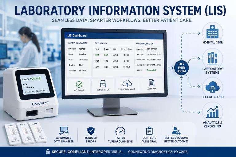
It cannot correct the binding chemistry. A hooked or interfered sample produces a wrong number that looks as precise as a right one, and the instrument adds its own variation from optics, strip positioning and temperature. Reader design is covered on the digital fluorescence reader page and software interpretation under AI-assisted signal analysis.
Where assay and materials collaborators fit
OncoFirm™ is developing a fluorescent lateral flow immunoassay platform and a prototype reader. Both are at the research stage, and their performance has not been established. The questions in this article are the open ones: hook and interference behavior in real clinical matrices, lot-to-lot reproducibility, and agreement with laboratory immunoassays.
Antibody and reagent developers, membrane suppliers and clinical chemistry laboratories can each take on part of that work. Current topics are listed under research and development priorities; to propose a joint project, use the research collaboration inquiry.
Frequently asked questions
What is the difference between a lateral flow assay and an ELISA?
Both use antibodies to capture and label a target. An ELISA runs in a plate with wash steps and enzyme signal amplification in a laboratory; a lateral flow assay runs in one step on a strip with no washing, which makes it simpler and usually less sensitive.
What is the hook effect in a lateral flow assay?
At very high target concentrations, excess target occupies the labeled and capture antibodies separately, so the test line signal drops and the sample can read falsely low. Dilution studies during validation show where this begins.
Are lateral flow assays quantitative?
Read by eye they are qualitative. With an instrument reader and lot-specific calibration they can report a concentration over a defined range, but that range and its precision have to be shown by validation.
Can a lateral flow assay detect cancer?
A strip measures a protein; it does not establish a diagnosis. Lateral flow assays for cancer-associated proteins are a research topic, and any result would need interpretation alongside imaging and pathology.
Related research and resources
- Fluorescent vs gold nanoparticle lateral flow assays — how the choice of label changes readout, quantitation and cost
- Point-of-care oncology testing — the evidence needed before a strip test is used near the patient
- Tumor antigen detection — the protein targets these assays are built to measure
- Fluorescent lateral flow platform — the research-stage platform OncoFirm is developing
References and further reading
- Posthuma-Trumpie GA, Korf J, van Amerongen A. Lateral flow (immuno)assay: its strengths, weaknesses, opportunities and threats. A literature survey. Anal Bioanal Chem. 2009;393(2):569–582.
- Sajid M, Kawde AN, Daud M. Designs, formats and applications of lateral flow assay: a literature review. J Saudi Chem Soc. 2015;19(6):689–705.
- Bahadır EB, Sezgintürk MK. Lateral flow assays: principles, designs and labels. TrAC Trends Anal Chem. 2016;82:286–306.
Research and regulatory status
OncoFirm™ technologies described on this site are in research and development. Their performance characteristics have not been established, and no OncoFirm product has been cleared, approved or authorized by the U.S. Food and Drug Administration. Nothing here is medical advice. Reference to a federal agency or funding program does not imply endorsement, funding, sponsorship or affiliation.