Liquid biopsy explained: analytes, workflow and early-stage limits
By OncoFirm Diagnostics · Last updated October 2026
A liquid biopsy is a test on blood or another body fluid that looks for material shed by a tumor: fragments of DNA, whole cells, vesicles or proteins. It gives molecular information without a tissue sample, which makes repeat testing practical, but it shows nothing of tissue architecture and does not replace pathology for diagnosis.
Clinical use is most established in advanced cancer, where circulating tumor DNA can identify targetable mutations when tissue is unavailable (Wan et al., 2017). Early detection is a harder problem, for reasons that are partly biological and partly statistical.
What a liquid biopsy measures
| Analyte | What it is | Typical methods | Main constraint |
|---|---|---|---|
| Cell-free DNA (cfDNA) | Short DNA fragments in plasma, mostly released by blood cells | Fragment profiling, methylation analysis, sequencing | Mostly non-tumor in origin |
| Circulating tumor DNA (ctDNA) | The tumor-derived fraction of cfDNA | Digital PCR; targeted or genome-wide sequencing | Can be a very small fraction, especially in early disease |
| Circulating tumor cells (CTCs) | Intact tumor cells in blood | Enrichment by size or surface markers, then imaging | Extremely rare among blood cells |
| Extracellular vesicles | Membrane-bound particles carrying protein and nucleic acid cargo | Isolation by centrifugation or affinity capture; cargo analysis | Isolation methods are not standardized |
| Proteins | Tumor-associated antigens and other circulating proteins | Immunoassay; mass spectrometry | Limited tumor specificity |
Summarized from the review literature (Heitzer et al., 2019).
From blood draw to report
The workflow begins before the laboratory. Blood cells lyse in the tube over time and release normal DNA that dilutes the tumor signal, so the collection tube, time to processing and centrifugation protocol all shape the result. Plasma is separated, usually in two spins, and cfDNA is extracted.
Analysis then depends on the question: digital PCR for a known mutation, targeted sequencing for a gene panel, or genome-wide methods for methylation and fragment patterns. Bioinformatic filtering separates true variants from sequencing error, and the report is read with imaging, pathology and clinical history.
Why early-stage disease is the hard case
Small tumors shed little DNA. A tube of blood holds a limited number of genome copies, so at very low tumor fractions the mutant fragment may simply be absent from the sample, a sampling limit that no gain in assay sensitivity can overcome (Wan et al., 2017).
Specificity has its own trap. Blood cells acquire mutations with age, a process called clonal hematopoiesis, and those variants appear in cfDNA where they can be mistaken for tumor signal (Heitzer et al., 2019).
The pattern shows in validation data: in a methylation-based multi-cancer test, sensitivity rose with stage (Klein et al., 2021). A prospective cohort study has also described the diagnostic workup that follows a positive signal (Schrag et al., 2023). The screening context is covered in why early cancer detection matters and where its limits lie.
Fragmentation patterns as a research direction
Mutations and methylation are not the only signals in cfDNA. Fragment sizes and their distribution across the genome differ between people with and without cancer, and machine-learning analysis of genome-wide fragmentation profiles is an active research area (Cristiano et al., 2019). As with any classifier, findings need validation in independent cohorts that include non-malignant conditions, such as inflammatory disease, which can also alter cfDNA.
How protein immunoassays relate to sequencing-based tests
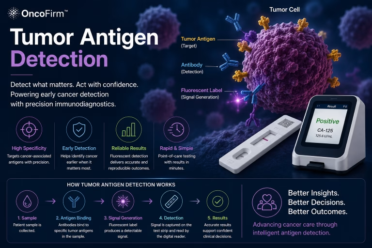
Proteins count as liquid biopsy analytes in the broad sense, but the trade-offs differ. Sequencing reads the tumor genome and can be highly specific, yet it needs centralized laboratories and bioinformatics. Protein immunoassays are generally cheaper, faster and run on simpler instruments, while the markers themselves are less tumor-specific, as tumor antigen detection explains.
The two are complementary more than competing. One multi-analyte study combined mutations in cfDNA with a panel of circulating proteins in a single test (Cohen et al., 2018). How the analyte classes compare is set out in what are cancer biomarkers.
Where research collaborators fit
OncoFirm™ is developing a research-stage fluorescent lateral flow immunoassay platform that targets proteins, not nucleic acids, and its performance has not been established. One open question is whether near-patient protein measurements add information to sequencing-based results when both are run on matched samples from the same patients.
That needs partners with paired plasma sets, staging data and ctDNA results: liquid biopsy laboratories, cancer center biobanks and biostatisticians. Related topics are listed under research and development priorities, and proposals go through the research collaboration inquiry.
Frequently asked questions
Does a liquid biopsy replace a tissue biopsy?
No. Tissue remains the standard for diagnosis because it shows histology and tumor architecture. Liquid biopsy adds molecular information and is most useful when tissue is unavailable or repeat sampling is needed.
What is the difference between cfDNA and ctDNA?
Cell-free DNA is all the fragmented DNA circulating in plasma, most of it from normal cells. Circulating tumor DNA is the portion that comes from tumor cells, and it can be a very small fraction of the total.
Can a liquid biopsy detect early-stage cancer?
Sometimes, but less reliably than advanced cancer, because small tumors release little DNA. Reported sensitivity in multi-cancer studies has been lowest for stage I disease.
Are protein tumor markers a form of liquid biopsy?
In the broad sense, yes: they are tumor-related analytes measured in blood. The term is more often used for circulating DNA and cells, which are analyzed by molecular methods and not by immunoassay.
Related research and resources
- Tumor antigen detection — the protein side of blood-based testing and its specificity limits
- Why early cancer detection matters — overdiagnosis, lead-time bias and false positives in screening
- Multi-biomarker blood tests for screening and triage — a research framework summary on evaluating multi-analyte tests
- AI in cancer diagnostics — machine learning in cancer diagnostics and how it is evaluated
References and further reading
- National Cancer Institute. Liquid biopsy — NCI Dictionary of Cancer Terms.
- Wan JCM, Massie C, Garcia-Corbacho J, et al. Liquid biopsies come of age: towards implementation of circulating tumour DNA. Nat Rev Cancer. 2017;17(4):223–238.
- Heitzer E, Haque IS, Roberts CES, Speicher MR. Current and future perspectives of liquid biopsies in genomics-driven oncology. Nat Rev Genet. 2019;20(2):71–88.
- Cohen JD, Li L, Wang Y, et al. Detection and localization of surgically resectable cancers with a multi-analyte blood test. Science. 2018;359(6378):926–930.
- Cristiano S, Leal A, Phallen J, et al. Genome-wide cell-free DNA fragmentation in patients with cancer. Nature. 2019;570(7761):385–389.
- Klein EA, Richards D, Cohn A, et al. Clinical validation of a targeted methylation-based multi-cancer early detection test using an independent validation set. Ann Oncol. 2021;32(9):1167–1177.
- Schrag D, Beer TM, McDonnell CH, et al. Blood-based tests for multicancer early detection (PATHFINDER): a prospective cohort study. Lancet. 2023;402(10409):1251–1260.
Research and regulatory status
OncoFirm™ technologies described on this site are in research and development. Their performance characteristics have not been established, and no OncoFirm product has been cleared, approved or authorized by the U.S. Food and Drug Administration. Nothing here is medical advice. Reference to a federal agency or funding program does not imply endorsement, funding, sponsorship or affiliation.